

doi: 10.1016/j.jneumeth.2008.05.007.
Epub 2008 May 20.
Single-synapse ablation and long-term imaging in live C. elegans
Affiliations
- PMID: 18579213
- PMCID: PMC2535809
- DOI: 10.1016/j.jneumeth.2008.05.007
Item in Clipboard
Single-synapse ablation and long-term imaging in live C. elegans
J Neurosci Methods.
.
Abstract
Synapses are individually operated, computational units for neural communication. To manipulate physically individual synapses in a living organism, we have developed a laser ablation technique for removing single synapses in live neurons in C. elegans that operates without apparent damage to the axon. As a complementary technique, we applied microfluidic immobilization of C. elegans to facilitate long-term fluorescence imaging and observation of neuronal development. With this technique, we directly demonstrated the existence of competition between developing synapses in the HSNL motor neuron.
Figures
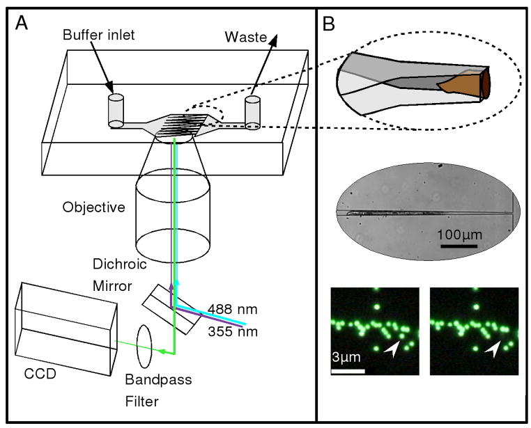
Overview of the experimental design. (A) Schematic illustrating the arrangement of the optical setup and microfluidic chip. Buffer flow is from left to right in the direction of the taper, such that worms are push towards the channel constrictions. Two laser sources, a blue (488 nm, CW) and an ultraviolet (355 nm, ~ 3ns pulse), are reflected from a dichroic mirror into the back aperture of a microscope objective lens and directed into the sample for fluorescence excitation and ablation, respectively. Fluorescence is collected and imaged with a CCD camera. (B) Top image: an illustration showing the details of the microfluidic system and the captured worm. The channels narrow to a final width of 8μm while maintaining a constant height of ~ 30μm. As a result, the worms tend to be rotated onto their side, as shown. Middle image: A bright-field image showing a worm trapped in the narrow region of the tapered channel. Bottom images: A before and after picture showing the selective ablation of a single 500 nm bead (indicated by arrow) in close proximity to other beads. The scale bar in the left image represents 3 μm.
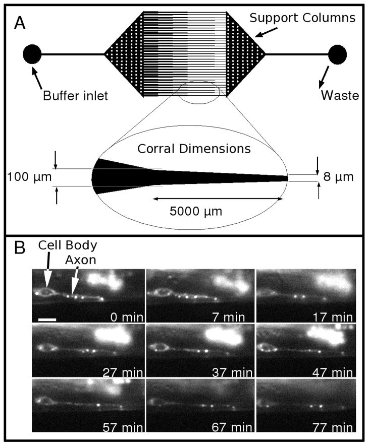
Details of the worm corral and sample data. (A) A CAD drawing of the photomask used to generate the master for the microfluidic chip. The inlet and outlet regions have circular posts (white dots) to prevent the elastomeric chip from collapsing. Inset below the CAD drawing shows details of the dimensions of the finely tapered region. (B) A series of fluorescence images from a time-lapse series showing the early development of the HSNL motor neuron expressing GFP::RAB-3 in a L4 stage worm immobilized in the microchannel. The animal was rotated onto its side in this image so the bend in the axon was along the Z axis. The scale bar in the 0 min frame represents 3 μm.
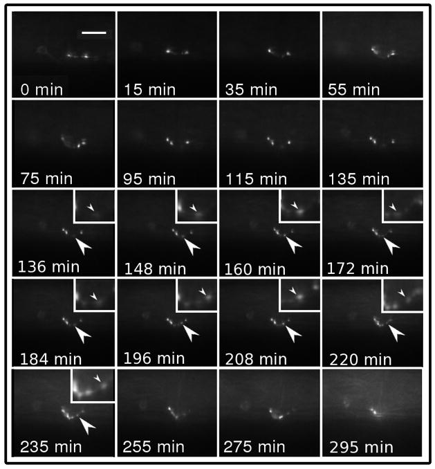
A series of fluorescence time-lapse images showing the development of the HSNL motor neuron expressing GFP::RAB-3 in a YA worm. The series shows the established morphology typical at the end of the L4 stage progress to a mature synapse pattern. In the full animated sequence, movement of material between the puncta is visible and no ectopic synapses can be seen to develop. The scale bar in the 0 min frame represents 6 μm.
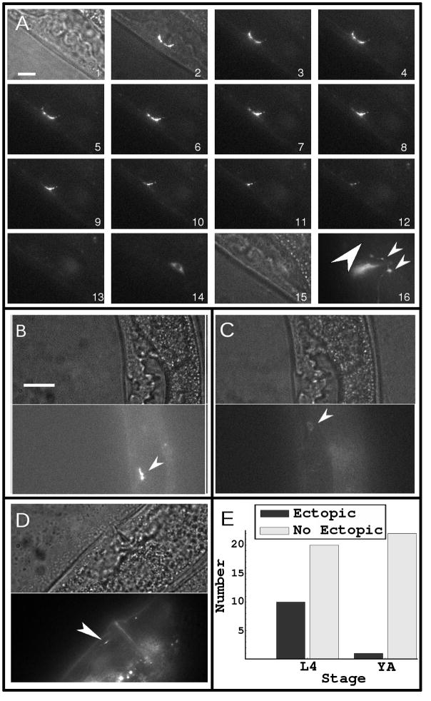
The results of the ablation experiments. (A) A sequence of images showing the ablation of a mature synapse made by the HSNL neuron. Because the size of this synapse was much larger than the focal spot of the UV laser, we typically had to use more than ten pulses to ablate the entire synapse. Frame 1 is a bright-field image and frame 2 is a merged bright-field and fluorescence image prior to the ablation of the synapse. Frames 3–13 show the synapse during ablation. Frame 14 shows the cell body, which was out of focus in frames 3–13. Frame 15 is a bright-field image of the region where the ablated synapse was, which showed no overt damage to the specimen. Frame 16 shows the same specimen as in frame 13 and 15, but after development for several hours, during which the synapses reappeared (small arrows). The large arrow points to the region where an ectopic synapse would have formed, which in this case did not. (B) shows another specimen at an earlier stage of development in bright field (top panel) and fluorescence (bottom panel) prior to ablation (the arrow indicates synaptic puncta). (C) shows the same specimen as in (B) after ablation in bright field (top panel) and fluorescence (bottom panel); the cell body can be seen in the fluorescence image and no overt damage is seen in the bright-field image (the arrow points to the cell body). (D) shows the same specimen as in (B) and (C) but after ablation and growth for several hours, after which an ectopic synapse had developed (arrow). (E) summarizes the results of 53 such experiments, which indicate ectopic synapses appear in worms in early developmental stage (L4) but not in YA worms. All scale bars represent 6 μm.
References
-
- Ding M, Chao D, Wang G, Shen K. Spatial Regulation of an E3 Ubiquitin Ligase Directs Selective Synapse Elimination. Science. 2007;317:947–51. - PubMed
-
- Duffy DC, McDonald JC, Schueller OJA, Whitesides GM. Rapid Prototyping of Microfluidic Systems in Poly(dimethylsiloxane) Anal Chem. 1998;70:4974–84. - PubMed
-
- Engert F, Bonhoeffer T. Synapse specificity of long-term potentiation breaks down at short distances. Nature. 1997;388:279–84. - PubMed
Publication types
MeSH terms
Grants and funding
LinkOut - more resources
Full Text Sources
Medical
Research Materials
