Review
. 2008 Nov 30;163(1-3):64-73.
doi: 10.1016/j.resp.2008.04.015. Epub 2008 Apr 30.

Respiratory impedance measurements for assessment of lung mechanics: focus on asthma

Affiliations
Review

Respiratory impedance measurements for assessment of lung mechanics: focus on asthma

Adam S LaPrad et al. Respir Physiol Neurobiol. .

Abstract

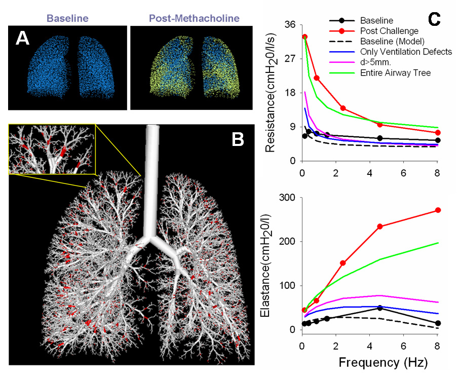
This review discusses the history and current state of the art of the forced oscillation technique (FOT) to measure respiratory impedance. We focus on how the FOT and its interaction with models have emerged as a powerful method to extract out not only clinically relevant information, but also to advance insight on the mechanisms and structures responsible for human lung diseases, especially asthma. We will first provide a short history of FOT for basic clinical assessment either directly from the data or in concert with lumped element models to extract out specific effective properties. We then spend several sections on the more exciting recent advances of FOT to probe the relative importance of tissue versus airway changes in disease, the impact of the disease on heterogeneous lung function, and the relative importance of small airways via synthesis of FOT with imaging. Most recently, the FOT approach has been able to directly probe airway caliber in humans and the distinct airway properties of asthmatics that seem to be required for airway hyperresponsiveness. We introduce and discuss the mechanism and clinical implications of this approach, which may be substantial for treatment assessment. Finally, we highlight important future directions for the FOT, particularly its use to probe specific lung components (e.g., isolated airways, isolated airway smooth muscle, etc.) and relate such data to the whole lung. The intent is to substantially advance an integrated understanding of structure-function relationships in the lung.


Figures

Figure 1
Figure 1
Two approaches to applying the FOT. (A) In the first technique, transfer impedance (Ztr) is measured by applying pressure oscillations around the chest wall and measuring flow at the airway opening. (B) In the second technique, input impedance (Zin) is measured by applying pressure oscillations at the airway opening and measuring flow at the airway opening. In both techniques, a high inertance bias tube (not shown) is used to allow the subject to breathe normally during forced oscillations of 4 Hz or greater (reproduced from Dubois et al., 1956 with permission).
Figure 2
Figure 2
The six-element lumped lung model. Airway resistance (Raw) and inertance (Iaw) are separated from tissue resistance (Rti), inertance (Iti), and compliance (Cti) with an alveolar gas compression term (Cg) (reproduced from Lutchen et al., 1993a with permission).
Figure 3
Figure 3
The real (Rrs) and imaginary (Xrs) parts of total lung impedance versus frequency for healthy subjects. Healthy subjects display a frequency dependent drop in Rrs. (reproduced from Hantos et al., 1986 with permission).
Figure 4
Figure 4
Relative contributions of airway resistance (dashed) and tissue resistance (solid) to lung resistance measurements obtained from parameter estimates of healthy human data. Raw is virtually the sole contributor to RL by 5 Hz (reproduced from Kaczka et al., 1997 with permission).
Figure 5
Figure 5
Measured lung mechanics [(A) lung resistance and (B) lung elastance] with the OVW technique for representative healthy, mild-to-moderate asthmatic, and severe asthmatic subjects. (A) With increasing severity of disease, RL becomes elevated and displays more frequency dependence. (B) Also, EL increases at higher frequencies due to airway wall shunting (adapted from Lutchen et al., 2001 with permission).
Figure 6
Figure 6
Simulated lung mechanics [(A) lung resistance and (B) lung elastance] from an airway tree model. Homogeneous peripheral constriction (narrowing all airways < 2mm diameter by 50%) results in a fairly uniform increase in RL and increased EL at higher frequencies due to airway wall shunting. However, heterogeneous peripheral constriction (20% mean, 50% coefficient of variation) results in elevated RL with increased frequency dependence of RL. Also, low frequency EL increases, indicative of airway closures (adapted from Lutchen et al., 2001 with permission).
Figure 7
Figure 7
Overview of the image functional modeling (IFM) approach. (A) The three-dimensional locations of the ventilation defects, defined from HP 3He RI images, are mapped into the alveolar terminal units from an anatomical airway tree model (shown in yellow). (B) Next, the largest airways are found that, when closed, will result in the same ventilation defects as the images without affecting other ventilated regions (shown in red). (C) Lastly, the constriction pattern is found to best match the measured respiratory mechanics. The necessary closed airways are consistently small (< 2 mm) and constrictions must be applied throughout the airway tree to match the lung mechanics.
Figure 8
Figure 8
Airway resistance plotted versus time for representative healthy and asthmatic subjects. After MCh challenge, Raw is much more elevated in the asthmatic subject compared to the typical healthy subject. However, when DIs are withheld before MCh challenge in the healthy subject, amplified reactivity to MCh occurs. Following MCh challenge, a single DI causes Raw to decrease to a minimum value (Rmin) at the peak of the DI (at TLC). Compared to the asthmatic subject, the healthy subject is able to reach a lower Rmin at the peak of the DI and remain at a lower Raw following DI, suggesting a limited bronchodilatory effect in asthmatic subjects (adapted from Black et al., 2004 with permission).
Figure 9
Figure 9
Dynamic lung elastance (EL) plotted versus time for a healthy subject (A) and an asthmatic subject (B). EL reflects a change in the fraction of lung tissue participating in breathing, where a decrease in EL would represent airway re-opening (or recruitment) and an increase would represent airway closure (or de-recruitment). In the asthmatic subject (B), EL increases after MCh challenge, but shows little change after DI. In the healthy subject (A), EL increases much greater when DIs are prohibited before MCh challenge. However, a DI after MCh challenge causes a substantially drop in EL. This suggests that the amplified reactivity that occurs in healthy subjects by prohibiting DIs is a consequence of heterogeneous airway closures and/or severe narrowings that occur simply by withholding DIs, but can be mostly ablated with a DI (reproduced from Black et al., 2004 with permission).
Figure 10
Figure 10
Airway conductance (mean linear regressions) plotted versus lung volume for healthy and asthmatic subjects before and after bronchodilator. Compared to healthy subjects, asthmatic subjects had significantly lower airway distensibility (slope of regressions) for lung volumes of 75–100% TLC. Also, administration of a bronchodilator had no effect in changing airway distensibility (reproduced from Brown et al., 2007 with permission).
Figure 11
Figure 11
Baseline Rmin plotted versus FEV1 for healthy and asthmatic subjects. The FEV1 values for asthmatic subjects vary greatly, making it impossible to distinguish asthmatic from healthy subjects by this measure. However, all asthmatic subjects had consistently higher baseline Rmin values compared to healthy subjects. Thus, it appears that a threshold in Rmin can be determined (dashed line), below which it would be highly improbable for a subject to be asthmatic (plotted from data partially presented in Black et al., 2004).
Figure 12
Figure 12
Multiple trajectories to AHR. The passive airway wall substructures and active airway smooth muscle compose the complete airway, both of which can be altered by the local inflammatory mediator environment. The airway network is embedded in a parenchymal tissue matrix, and combined these components constitute the whole lung. Thus, any, all, or a combination of components can be responsible for AHR and altered lung function measurable with FOT.

References

    1. Avanzolini G, Barbini P, Cappello A, Cevenini G. Real-time tracking of parameters of lung mechanics: emphasis on algorithm tuning. J. Biomed. Eng. 1990;12:489–495. - PubMed
    1. Bates JH, Lauzon AM, Dechman GS, Maksym GN, Schuessler TF. Temporal dynamics of pulmonary response to intravenous histamine in dogs: effects of dose and lung volume. J. Appl. Physiol. 1994;76:616–626. - PubMed
    1. Bates JH, Ludwig MS, Sly PD, Brown K, Martin JG, Fredberg JJ. Interrupter resistance elucidated by alveolar pressure measurement in open-chest normal dogs. J. Appl. Physiol. 1988;65:408–414. - PubMed
    1. Bates JH, Lutchen KR. The interface between measurement and modeling of peripheral lung mechanics. Respir. Physiol. Neurobiol. 2005;148:153–164. - PubMed
    1. Black LD, Dellaca R, Jung K, Atileh H, Israel E, Ingenito EP, Lutchen KR. Tracking variations in airway caliber by using total respiratory vs. airway resistance in healthy and asthmatic subjects. J. Appl. Physiol. 2003;95:511–518. - PubMed