

Regulation of the Helicobacter pylori cellular receptor decay-accelerating factor
- PMID: 18579524
- PMCID: PMC2527108
- DOI: 10.1074/jbc.M801144200
Regulation of the Helicobacter pylori cellular receptor decay-accelerating factor
Abstract
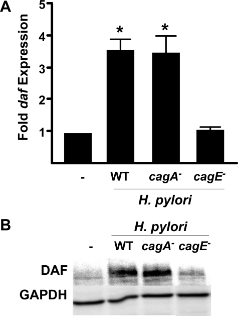
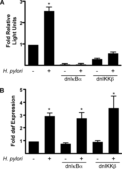
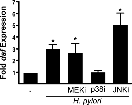
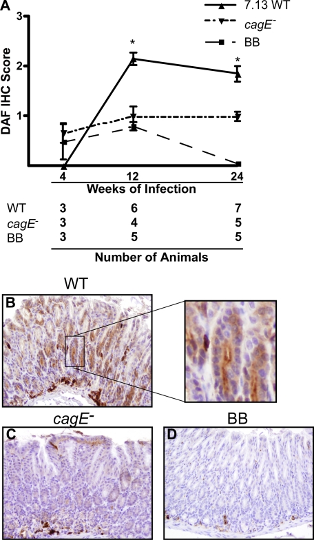
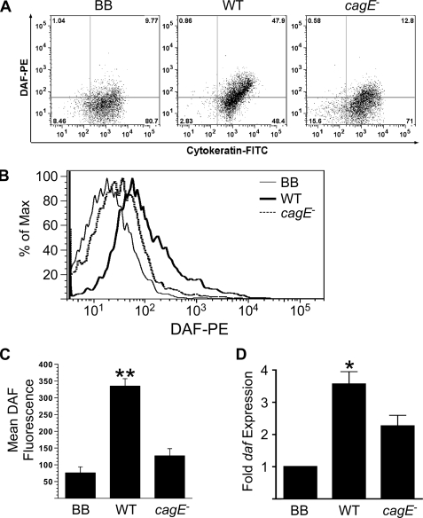
Chronic gastritis induced by Helicobacter pylori is the strongest known risk factor for peptic ulceration and distal gastric cancer, and adherence of H. pylori to gastric epithelial cells is critical for induction of inflammation. One H. pylori constituent that increases disease risk is the cag pathogenicity island, which encodes a secretion system that translocates bacterial effector molecules into host cells. Decay-accelerating factor (DAF) is a cellular receptor for H. pylori and a mediator of the inflammatory response to this pathogen. H. pylori induces DAF expression in human gastric epithelial cells; therefore, we sought to define the mechanism by which H. pylori up-regulates DAF and to extend these findings into a murine model of H. pylori-induced injury. Co-culture of MKN28 gastric epithelial cells with the wild-type H. pylori cag(+) strain J166 induced transcriptional expression of DAF, which was attenuated by disruption of a structural component of the cag secretion system (cagE). H. pylori-induced expression of DAF was dependent upon activation of the p38 mitogen-activated protein kinase pathway but not NF-kappaB. Hypergastrinemic INS-GAS mice infected with wild-type H. pylori demonstrated significantly increased DAF expression in gastric epithelium versus uninfected controls or mice infected with an H. pylori cagE(-) isogenic mutant strain. These results indicate that H. pylori cag(+) strains induce up-regulation of a cognate cellular receptor in vitro and in vivo in a cag-dependent manner, representing the first evidence of regulation of an H. pylori host receptor by the cag pathogenicity island.
Figures
Similar articles
-
Helicobacter pylori promotes the expression of Krüppel-like factor 5, a mediator of carcinogenesis, in vitro and in vivo.PLoS One. 2013;8(1):e54344. doi: 10.1371/journal.pone.0054344. Epub 2013 Jan 23. PLoS One. 2013. PMID: 23372710 Free PMC article.
-
Helicobacter pylori Depletes Cholesterol in Gastric Glands to Prevent Interferon Gamma Signaling and Escape the Inflammatory Response.Gastroenterology. 2018 Apr;154(5):1391-1404.e9. doi: 10.1053/j.gastro.2017.12.008. Epub 2017 Dec 19. Gastroenterology. 2018. PMID: 29273450
-
Helicobacter pylori strain-selective induction of matrix metalloproteinase-7 in vitro and within gastric mucosa.Gastroenterology. 2003 Oct;125(4):1125-36. doi: 10.1016/s0016-5085(03)01206-x. Gastroenterology. 2003. PMID: 14517796
-
Pathogenicity island-dependent effects of Helicobacter pylori on intracellular signal transduction in epithelial cells.Int J Med Microbiol. 2005 Sep;295(5):335-41. doi: 10.1016/j.ijmm.2005.06.007. Int J Med Microbiol. 2005. PMID: 16173500 Review.
-
Molecular response of gastric epithelial cells to Helicobacter pylori-induced cell damage.Cell Microbiol. 1999 Sep;1(2):93-9. doi: 10.1046/j.1462-5822.1999.00018.x. Cell Microbiol. 1999. PMID: 11207544 Review.
Cited by
-
Helicobacter pylori promotes the expression of Krüppel-like factor 5, a mediator of carcinogenesis, in vitro and in vivo.PLoS One. 2013;8(1):e54344. doi: 10.1371/journal.pone.0054344. Epub 2013 Jan 23. PLoS One. 2013. PMID: 23372710 Free PMC article.
-
The active form of Helicobacter pylori vacuolating cytotoxin induces decay-accelerating factor CD55 in association with intestinal metaplasia in the human gastric mucosa.J Pathol. 2022 Oct;258(2):199-209. doi: 10.1002/path.5990. Epub 2022 Aug 18. J Pathol. 2022. PMID: 35851954 Free PMC article.
-
PI3K/Akt pathway restricts epithelial adhesion of Dr + Escherichia coli by down-regulating the expression of decay accelerating factor.Exp Biol Med (Maywood). 2014 May;239(5):581-94. doi: 10.1177/1535370214522183. Epub 2014 Mar 5. Exp Biol Med (Maywood). 2014. PMID: 24599886 Free PMC article.
-
Helicobacter infection alters MyD88 and Trif signalling in response to intestinal ischaemia-reperfusion.Exp Physiol. 2011 Feb;96(2):104-13. doi: 10.1113/expphysiol.2010.055426. Epub 2010 Nov 5. Exp Physiol. 2011. PMID: 21056969 Free PMC article.
-
Sex-specific signaling through Toll-Like Receptors 2 and 4 contributes to survival outcome of Coxsackievirus B3 infection in C57Bl/6 mice.Biol Sex Differ. 2012 Dec 15;3(1):25. doi: 10.1186/2042-6410-3-25. Biol Sex Differ. 2012. PMID: 23241283 Free PMC article.
References
-
- Peek, R. M., Jr., and Blaser, M. J. (2002) Nat. Rev. Cancer 2 28-37 - PubMed
-
- Moss, S. F., and Sood, S. (2003) Curr. Opin. Infect. Dis. 16 445-451 - PubMed
-
- Moss, S. F., and Blaser, M. J. (2005) Nat. Clin. Pract. Oncol. 2 90-97 - PubMed
-
- Blanchard, T. G., Drakes, M. L., and Czinn, S. J. (2004) Curr. Opin. Gastroenterol. 20 10-15 - PubMed
Publication types
MeSH terms
Substances
Grants and funding
LinkOut - more resources
Full Text Sources
Medical
Molecular Biology Databases
Miscellaneous
