. 2008 Aug 29;283(35):23914-21.
doi: 10.1074/jbc.M800776200. Epub 2008 Jun 25.

Analgesic compound from sea anemone Heteractis crispa is the first polypeptide inhibitor of vanilloid receptor 1 (TRPV1)

Affiliations

Analgesic compound from sea anemone Heteractis crispa is the first polypeptide inhibitor of vanilloid receptor 1 (TRPV1)

Yaroslav A Andreev et al. J Biol Chem. .

Abstract

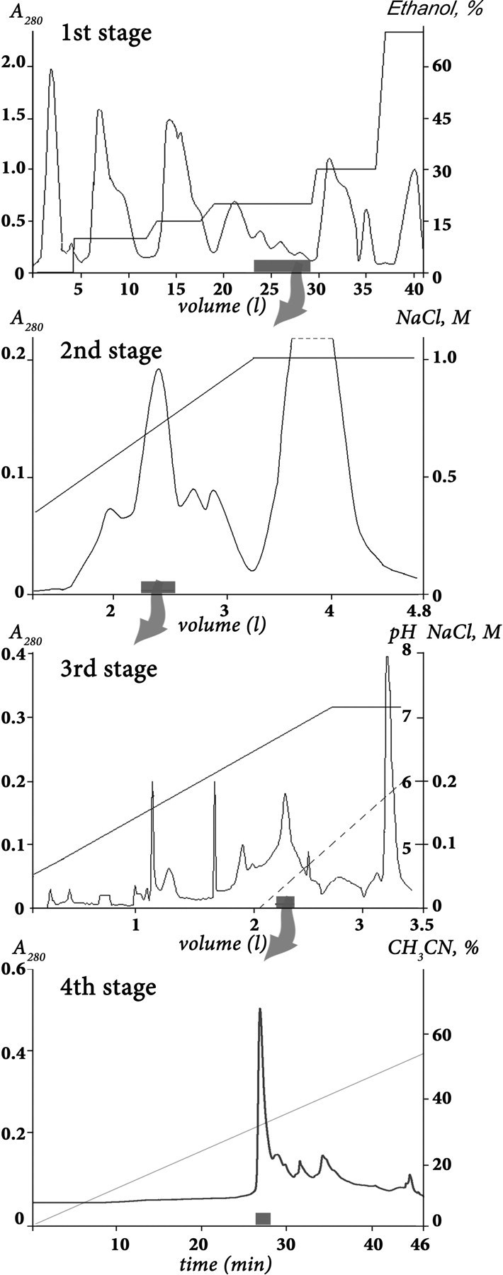
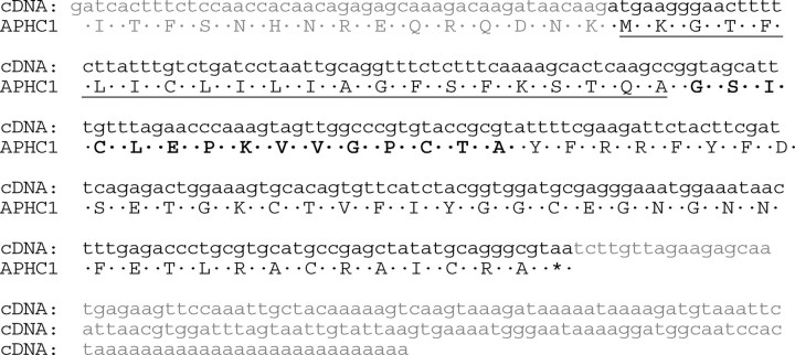
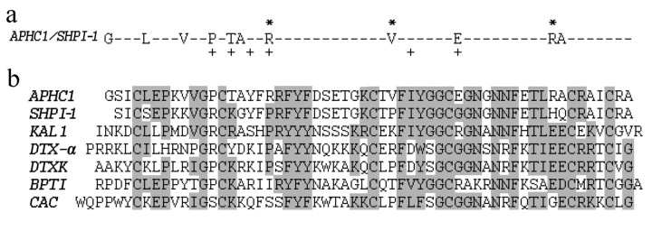
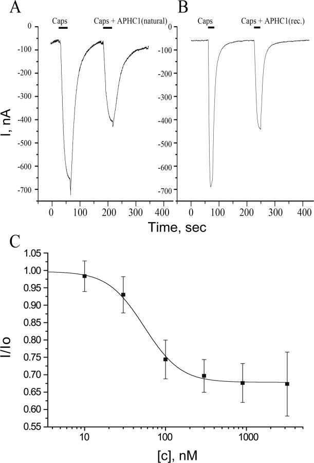
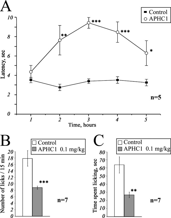
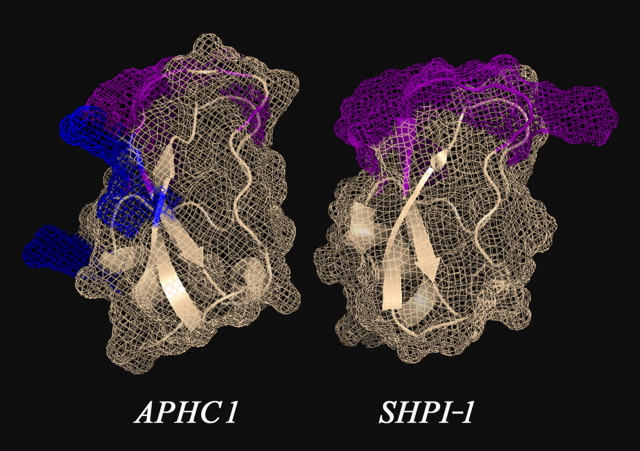
Venomous animals from distinct phyla such as spiders, scorpions, snakes, cone snails, or sea anemones produce small toxic proteins interacting with a variety of cell targets. Their bites often cause pain. One of the ways of pain generation is the activation of TRPV1 channels. Screening of 30 different venoms from spiders and sea anemones for modulation of TRPV1 activity revealed inhibitors in tropical sea anemone Heteractis crispa venom. Several separation steps resulted in isolation of an inhibiting compound. This is a 56-residue-long polypeptide named APHC1 that has a Bos taurus trypsin inhibitor (BPTI)/Kunitz-type fold, mostly represented by serine protease inhibitors and ion channel blockers. APHC1 acted as a partial antagonist of capsaicin-induced currents (32 +/- 9% inhibition) with half-maximal effective concentration (EC(50)) 54 +/- 4 nm. In vivo, a 0.1 mg/kg dose of APHC1 significantly prolonged tail-flick latency and reduced capsaicin-induced acute pain. Therefore, our results can make an important contribution to the research into molecular mechanisms of TRPV1 modulation and help to solve the problem of overactivity of this receptor during a number of pathological processes in the organism.


Figures

FIGURE 1.
FIGURE 1.
Purification of APHC1. The first separation stage of a dried ethanol extract of sea anemone H. crispa nematocysts was done on a water-equilibrated hydrophobic column Polychrom-1 (7 × 30 cm). Fractions were eluted by stepwise ethanol gradient with a flow rate of 1.2 liters/h. Active fraction (marked as a gray box on overall separation steps) has been separated on the second stage by ion exchange chromatography on Bio-Rex 70 column (2.5 × 60 cm). The separation was done in 5 mm ammonium acetate buffer (pH 4.5) by flow rate 22 ml/h in a linear gradient of NaCl concentration. The third stage of purification was performed with a flow rate 70 ml/h on the ion exchange column SP-Sephadex C-25 (2.5 × 40 cm), with the same 5 mm ammonium acetate buffer (start buffer, pH 4.5) in combined gradient of NaCl concentration and pH value. Final purification (stage 4) was achieved on a reverse-phase column Jupiter C5 (4.6 × 150 mm) in 0.1% trifluoroacetic acid with a flow rate of 1 ml/min using a linear gradient of acetonitrile concentration.
FIGURE 2.
FIGURE 2.
Structure of APHC1 gene determined from cDNA clones sequences. The signal peptide sequence deduced in the precursor structure is underlined, and partial N-terminal fragment determined by Edman degradation is shown in bold.
FIGURE 3.
FIGURE 3.
Alignment of APHC1 primary structure and BPTI/Kunitz type polypeptides. a, substitution plot of pairwise residues among APHC1 and sea anemone trypsin inhibitor SHPI-1 from S. helianthus (21). Amino acid residues essential for inhibitory activity of SHPI-1 as described in Ref. are marked with a plus sign, and arginine/valine residues that are probably important in APHC1 are marked with asterisks. b, multiple alignment of APHC1 amino acid sequence with serine protease inhibitor SHPI-1 from sea anemone S. helianthus (21); kalicludine 1 (KAL1), K+ channel and trypsin inhibitor from sea anemone Anemonia sulcata (23); two dendrotoxins active on K+ channel DTX α from snake D. angusticeps (24) and DTXK from Dendroaspis polylepis polylepis (25); BPTI (26); and calcicludine (CAC), a blocker of high threshold Ca2+ channels from snake D. angusticeps (27). Related or similar residues are shadowed.
FIGURE 4.
FIGURE 4.
Action of APHC1 on TRPV1 channels. A and B, application traces of capsaicin (Caps) alone and capsaicin mixed with purified natural APHC1 (final concentration 500 nm) (A) or with recombinant (rec.) APHC1 (final concentration 300 nm) (B). Channels were activated by 2 μm capsaicin 35 s (A) or 20 s (B) before washing, and oocytes were held at -50 mV. C, dose-response curve for APHC1 inhibitor activity on capsaicin-activated TRPV1 channels. The abscissa axis represents the ratio of ion current evoked by co-application of agonist and APHC1 (I) to ion current evoked by agonist alone (Io) on the same oocyte. Each point represents the mean ± S.E. with n = 4–8.
FIGURE 5.
FIGURE 5.
Analgesic (antinociception) effect of a recombinant APHC1. A, tail-flick test. Latency indicates the delay of tail-flick response after one intramuscular administration of APHC1 (0.1 mg/kg) into wild-type mice, n = 5. B and C, attenuation of mice capsaicin-induced behavior by intravenous administered APHC1 (0.1 mg/kg), n = 7. Results are presented as mean ± S.E., ***, p < 0.005, **, p < 0.01, *, p < 0.06 versus control (analysis of variance and Tukey's test).
FIGURE 6.
FIGURE 6.
Three-dimensional structural models of sea anemones polypeptides. The structure of SHPI-1-trypsin inhibitor from sea anemone S. helianthus (Protein Data Bank ID 1SHP) is shown. The spatial structure of APHC1 was calculated from SHPI-1 data by molecular modeling. Amino acid residues essential for serine protease inhibition are painted in magenta on both models, and two arginine residues (Arg-18 and Arg-48) and one valine residue (Val-31), presumably important for APHC1 activity, are shown in blue.

References

    1. Kawai, N., and Nakajima, T. (1993) in Natural and Synthetic Neurotoxins (Harvey, A. L., ed), pp. 319-345, Academic Press, London
    1. Miljanich, G. P. (1997) Sci. Med. (Phila.) 4 6-15
    1. Mortaria, M. R., Cunhaa, A. O., Ferreiraa, L. B., and Ferreira dos Santos, W. (2007) Pharmacol. Ther. 114 171-183 - PubMed
    1. Watters, M. R. (2005) Semin. Neurol. 25 278-289 - PubMed
    1. Guo, A., Vulchanova, L., Wang, J., Li, X., and Elde, R. (1999) Eur. J. Neurosci. 11 946-958 - PubMed

Publication types

MeSH terms

Associated data

LinkOut - more resources