. 2008 Sep;82(17):8863-70.
doi: 10.1128/JVI.00343-08. Epub 2008 Jun 25.

Association of respiratory syncytial virus M protein with viral nucleocapsids is mediated by the M2-1 protein

Affiliations

Association of respiratory syncytial virus M protein with viral nucleocapsids is mediated by the M2-1 protein

Dongsheng Li et al. J Virol. 2008 Sep.

Abstract

Cytoplasmic inclusions in respiratory syncytial virus-infected cells comprising viral nucleocapsid proteins (L, N, P, and M2-1) and the viral genome are sites of viral transcription. Although not believed to be necessary for transcription, the matrix (M) protein is also present in these inclusions, and we have previously shown that M inhibits viral transcription. In this study, we have investigated the mechanisms for the association of the M protein with cytoplasmic inclusions. Our data demonstrate for the first time that the M protein associates with cytoplasmic inclusions via an interaction with the M2-1 protein. The M protein colocalizes with M2-1 in the cytoplasm of cells expressing only the M and M2-1 proteins and directly interacts with M2-1 in a cell-free binding assay. Using a cotransfection system, we confirmed that the N and P proteins are sufficient to form cytoplasmic inclusions and that M2-1 localizes to these inclusions; additionally, we show that M associates with cytoplasmic inclusions only in the presence of the M2-1 protein. Using truncated mutants, we show that the N-terminal 110 amino acids of M mediate the interaction with M2-1 and the subsequent association with nucleocapsids. The interaction of M2-1 with M and, in particular, the N-terminal region of M may represent a target for novel antivirals that block the association of M with nucleocapsids, thereby inhibiting virus assembly.


Figures
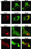
FIG. 1.
FIG. 1.
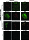
The M protein is present in cytoplasmic inclusions in RSV-infected cells. RSV-infected HEp2 cells were fixed 18 h after infection and were double stained with various antibody combinations, followed by CLSM analysis. The antibodies used are indicated above each panel. All primary antibodies were used at a dilution of 1/100 in PBS and incubated for 30 min; cells were washed with PBS and incubated for 30 min with species-specific secondary antibodies conjugated to fluorescein isothiocyanate or Texas Red. Cells were washed again, and localization was observed using CLSM. The left and middle columns of images are the red and green channel outputs of the same cell at the same optical plane; the right columns of images are computer-generated, merged images of the two channels, with yellow coloration indicating colocalization. A rabbit anti-M antiserum was used in C, and a mouse anti-M antibody was used in D.
FIG. 2.
FIG. 2.
Number of M-containing cytoplasmic inclusions increases over time. RSV-infected HEp2 cells were fixed at 14 h, 16 h, and 20 h postinfection and double stained with a mouse anti-M antibody and guinea pig anti-M2-1 antiserum, followed by CLSM, as described in legend of Fig. 1. Numbers on the right indicate proportions of inclusions containing M2-1 that also contain M [(number of inclusions containing M)/(number of inclusions containing M2-1)]; data from ≥20 cells are presented (means ± standard errors).
FIG. 3.
FIG. 3.
M interacts directly with the M2-1 protein. (A) HA epitope-tagged M mRNA was transfected into HEp2 cells along with SFV-M2-1 (top), SFV-N (middle), or SFV-P mRNAs (bottom). The cells were fixed 15 h after transfection, followed by double staining with the indicated antibodies and CLSM analysis. (B) Five microliters of cell lysate from nontransfected cells and cells transfected to express M2-1-HA, P-HA, and N-HA was resolved by 12% SDS-PAGE, followed by Western blotting using rabbit anti-HA antibody. (C and D) Equivalent amounts of M2-1-HA, P-HA, and P-HA (as estimated from the Western blot) and a similar volume of untransfected cell lysate were diluted and incubated with bacterially expressed M (C) or hepatitis C virus NS3 (D) immobilized on microtiter plate wells. Bound protein was detected with rabbit anti-HA antibody followed by horseradish peroxidase-conjugated secondary antibody and development with tetramethyl benzidine substrate.
FIG. 4.
FIG. 4.
M associates with inclusions only in the presence of M2-1. N and P mRNAs were cotransfected into HEp2 cells with M2-1 (top row), M-HA (second row), or M2-1 and M-HA mRNAs (third row). Control (mock) cells were transfected with RNA from an empty SFV replicon (bottom row). Cells were fixed at 15 h after transfection and double stained with the indicated antibodies, and fluorescence was visualized using CLSM.
FIG. 5.
FIG. 5.
The N terminus of M appears to mediate associations with inclusions and binding to M2-1. (A) Schematic diagram showing constructs of M used. The sequence motifs are based on predictive software analysis of the amino acid sequence and our previous work. NES, nuclear export signal; NLS, nuclear localization signal; LRR, leucine-rich region. Numbers indicate amino acids within the M sequence; each construct has a C-terminal HA tag. Broken lines in the mutants indicate the actual boundaries of the ZFD in the full-length protein. Full-length M and all three mutants were localized throughout the cell, as shown by the respective CLSM images on the right. (B) SFV-N, SFV-P, and SFV-M2-1 mRNAs were cotransfected into HEp2 cells with M-HA, M-ΔN-HA, M-ΔC-HA, or M-Δ(C+ZFD)-HA mRNA, and cells were fixed 15 h after transfection, followed by immunofluorescence assays using anti-HA and anti-P antibodies, as indicated, and CLSM analysis. Arrowheads point to cytoplasmic inclusions that have M (or its mutant). Numbers on the right indicate the proportion of inclusions containing P that also contain M [(number of inclusions containing M)/(number of inclusions containing P)] (means ± standard errors). (C) M-HA, M-ΔN-HA, M-ΔC-HA, M-Δ(C+ZFD)-HA, and luciferase were translated in RRL from in vitro-transcribed mRNAs. Five microliters of each translation reaction mixture was resolved by 12% SDS-PAGE, followed by immunoblotting using rabbit anti-HA antibody. (D) Equivalent amounts of translated M-HA, M-ΔN-HA, M-ΔC-HA, M-Δ(C+ZFD)-HA, and luciferase were incubated with same amounts of lysates of cells expressing M2-1. Another control consisted of M-HA incubated with untransfected (mock) cell lysate. The mixtures were immunoprecipitated using anti-M2-1 antibody, and bound M-HA (or mutants) in the immunoprecipitate was eluted and detected by Western blotting using rabbit anti-HA antibody. (E) Densities of specific M-HA (or mutants) bands from Western blots like those shown in C and D were measured using a Molecular Imager system (Bio-Rad). The percentage of total protein recovered is shown. The data represent means ± standard deviations of data from three independent experiments.

References

    1. Carromeu, C., F. M. Simabuco, R. E. Tamura, L. E. F. Arcieri, and A. M. Ventura. 2007. Intracellular localization of human respiratory syncytial virus L protein. Arch. Virol. 1522259-2263. - PubMed
    1. Cathomen, T., B. Mrkic, D. Spehner, R. Drillien, R. Naef, J. Pavlovic, A. Aguzzi, M. A. Billeter, and R. Cattaneo. 1998. A matrix-less measles virus is infectious and elicits extensive cell fusion: consequences for propagation in the brain. EMBO J. 173899-3908. - PMC - PubMed
    1. Collins, P. L., R. M. Chanock, and B. R. Murphy. 2001. Respiratory syncytial viruses, p. 1443-1485. In D. M. Knipe and P. M. Howley (ed.), Fields virology, 4th ed., vol. 1. Lippincott Williams & Wilkins, Philadelphia, PA.
    1. Collins, P. L., M. G. Hill, J. Cristina, and H. Grosfeld. 1996. Transcription elongation factor of respiratory syncytial virus, a nonsegmented negative-strand RNA virus. Proc. Natl. Acad. Sci. USA 9381-85. - PMC - PubMed
    1. Coronel, E. C., K. G. Murti, T. Takimoto, and A. Portner. 1999. Human parainfluenza virus type 1 matrix and nucleoprotein genes transiently expressed in mammalian cells induce the release of virus-like particles containing nucleocapsid-like structures. J. Virol. 737035-7038. - PMC - PubMed

Publication types

Substances

LinkOut - more resources