. 2008 Sep;82(17):8797-811.
doi: 10.1128/JVI.00592-08. Epub 2008 Jun 25.

Protein kinase A-dependent step(s) in hepatitis C virus entry and infectivity

Affiliations

Protein kinase A-dependent step(s) in hepatitis C virus entry and infectivity

Michelle J Farquhar et al. J Virol. 2008 Sep.

Abstract

Viruses exploit signaling pathways to their advantage during multiple stages of their life cycle. We demonstrate a role for protein kinase A (PKA) in the hepatitis C virus (HCV) life cycle. The inhibition of PKA with H89, cyclic AMP (cAMP) antagonists, or the protein kinase inhibitor peptide reduced HCV entry into Huh-7.5 hepatoma cells. Bioluminescence resonance energy transfer methodology allowed us to investigate the PKA isoform specificity of the cAMP antagonists in Huh-7.5 cells, suggesting a role for PKA type II in HCV internalization. Since viral entry is dependent on the host cell expression of CD81, scavenger receptor BI, and claudin-1 (CLDN1), we studied the role of PKA in regulating viral receptor localization by confocal imaging and fluorescence resonance energy transfer (FRET) analysis. Inhibiting PKA activity in Huh-7.5 cells induced a reorganization of CLDN1 from the plasma membrane to an intracellular vesicular location(s) and disrupted FRET between CLDN1 and CD81, demonstrating the importance of CLDN1 expression at the plasma membrane for viral receptor activity. Inhibiting PKA activity in Huh-7.5 cells reduced the infectivity of extracellular virus without modulating the level of cell-free HCV RNA, suggesting that particle secretion was not affected but that specific infectivity was reduced. Viral particles released from H89-treated cells displayed the same range of buoyant densities as did those from control cells, suggesting that viral protein association with lipoproteins is not regulated by PKA. HCV infection of Huh-7.5 cells increased cAMP levels and phosphorylated PKA substrates, supporting a model where infection activates PKA in a cAMP-dependent manner to promote virus release and transmission.


Figures

FIG. 1.
FIG. 1.
Inhibition of PKA attenuates HCV infection. (A) Huh-7.5 cells were incubated with Ro-31-8220 (1 μM) (PKC inhibitor), H89 (10 μM) (PKA inhibitor), PD98059 (10 μM) (MEK1 inhibitor), U0126 (10 μM) (MEK1/2 inhibitor), or SB203580 (10 μM) (p38MEK inhibitor) for 1 h and infected with JFH-1 for 1 h in the presence of inhibitors. (B) Dose-dependent reduction of HCV infection by H89 inhibition of PKA. Huh-7.5 cells were incubated for 1 h with increasing concentrations of H89 and infected with J6/JFH (□) or JFH-1 (▵). (C) Dose-dependent reduction of HCV infection by myrPKI inhibition of PKA. Huh-7.5 cells were incubated for 1 h with increasing concentrations of myrPKI and infected with J6/JFH (□) or JFH-1 (▵). Infectivity is expressed relative to untreated control cells and represents the mean of three replicate infections. The data presented are from a single experiment and are representative of three independent experiments.
FIG. 2.
FIG. 2.
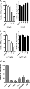
PKA-dependent HCV entry. (A) Dose-dependent reduction of HCVpp entry by H89 inhibition of PKA. Huh-7.5 cells were incubated for 1 h with increasing concentrations of H89 and infected with HCVpp-JFH-1 (white bars) or MLVpp (black bars). (B) Dose-dependent reduction of HCVpp entry by myrPKI inhibition of PKA. Huh-7.5 cells were incubated for 1 h with increasing concentrations of myrPKI and infected with HCVpp-H77 (white bars) or MLVpp (black bars). (C) Huh-7.5, Hep3B, Huh-7, 293T-CLDN1, and primary human hepatocytes (PHH) were incubated with H89 (10 μM) for 1 h and infected with HCVpp-H77. HCVpp-JFH-1 infection was assessed by flow cytometry, while for HCVpp-H77 infection, the cells were lysed, and the mean luciferase activity (relative light units) was measured; for all samples, the average Envpp value was subtracted. Infectivity is expressed relative to those of untreated control cells and represents the mean of data from three replicate infections. The data presented are from a single experiment and are representative of three independent experiments.
FIG. 3.
FIG. 3.
Putative role for PKAII in HCV infection. (A) Huh-7.5 cells were incubated with the cAMP analogs Rp-cAMPS (500 μM) or Rp-8-Br-cAMPS (500 μM) and infected for 1 h with J6/JFH (white bars) or JFH-1 (black bars). HCVcc-infected cells were fixed after 48 h and stained for NS5A, and the mean number of infected cells per well was determined by flow cytometry. Infectivity is expressed relative to untreated control cells and represents the mean of data from three replicate infections. The data presented are from a single experiment and are representative of three independent experiments. (B) Huh-7.5 cells expressing PKAIα (white bars) or PKAIIα (black bars) sensors were preincubated with Rp-cAMPS (500 μM) or Rp-8-Br-cAMPS (500 μM) for 1 h, followed by FK/IBMX (50 μM and 500 μM, respectively) stimulation for 30 min. The BRET signal was quantified using a Fusionα FP microplate reader, and data are plotted relative to untreated control cells. The data were compiled from three independent experiments. (C) Huh-7.5 cells expressing PKAIα (white bars) or PKAIIα (black bars) sensors were preincubated with Rp-cAMPS (500 μM) or Rp-8-Br-cAMPS (500 μM) for 1 h, followed by FK (10 μM) stimulation for 30 min. The data presented are compiled from three independent experiments. Statistical analysis using a Newman-Keuls multiple-comparison test confirms that FK treatment significantly reduced the BRET signal compared to those of control untreated cells (P < 0.05), while pretreatment with Rp-cAMPS had no significant effect on the BRET signal relative to the control. Preincubation of cells with Rp-8-Br-cAMPS did not inhibit the FK-stimulated decrease in BRET signal compared to the control.
FIG. 4.
FIG. 4.
FK activation of PKA enhances HCV infection. Huh-7.5 cells were incubated with FK (10 μM) (PKA activator) for 1 h with or without a 1-h preincubation with H89 (10 μM) (PKA inhibitor). Cells were infected with J6/JFH (white bars) or JFH-1 (black bars) (A) and HCVpp-JFH (B) for 1 h in the presence of PKA modulators. HCVcc-infected cells were fixed after 48 h and stained for NS5A, and the mean number of infected cells per well was determined by flow cytometry. For HCVpp, the cells were lysed, the mean luciferase activity (relative light units) was measured, and the average Envpp value was subtracted. Infectivity is expressed relative to untreated control cells and represents the mean of data for three replicate infections. The data presented are from a single experiment and are representative of three independent experiments.
FIG. 5.
FIG. 5.
Inhibition of PKA disrupts CLDN1 localization in Huh-7.5 cells. (A) Huh-7.5 cells were seeded onto glass coverslips and incubated with dimethyl sulfoxide (DMSO) (control), H89 (10 μM), FK (10 μM), Rp-cAMPS (500 μM), or Rp-8-Br-cAMPS (500 μM). Cells were fixed and stained for CLDN1 or CD81. (B) Huh-7.5 cells were incubated with DMSO (control) or H89 (10 μM) for 1 h, fixed, and stained for the TJ proteins ZO-1 and occludin. Bound antibodies were visualized using Alexa Fluor 488 anti-mouse Ig. (C) Huh-7.5 cells were transduced to express GFP-AKAP-IS or GFP-Scrambled peptide (green) and stained for CLDN1. Bound antibodies were visualized using Alexa Fluor 633 anti-mouse antibodies (red). Nuclei were visualized using DAPI (blue). Laser scanning confocal microscopic images of single 1-μm z sections were obtained using a 63× 1.2-numerical-aperture objective (scale bar represents 10 μm).
FIG. 6.
FIG. 6.
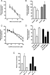
PKA-dependent infectivity of extracellular HCV. (A) Dose-dependent reduction in extracellular HCV infectivity by H89. J6/JFH (□)- and JFH-1 (▵)-infected Huh-7.5 cells were seeded in 48-well plates and incubated with increasing concentrations of H89 the following day. Cells were extensively washed, supernatant was collected after 1 h, and infectivity was quantified. (B) J6/JFH-infected cells were incubated with H89 (10 μM) for 1 h (white bars) or 8 h (gray bars), the intracellular virus was released by repeated freeze-thaw cycles, and infectivity was quantified. (C) Dose-dependent reduction in extracellular HCV infectivity by myrPKI. J6/JFH (□)- and JFH-1 (▵)-infected Huh-7.5 cells were seeded in 48-well plates and incubated with increasing concentrations of myrPKI the following day. Cells were extensively washed, supernatant was collected after 1 h, and infectivity was quantified. (D) The infectivity of extracellular virus obtained from J6/JFH (white bars)- and JFH-1 (black bars)-infected Huh-7.5 cells incubated with the isoform-specific PKA inhibitors Rp-cAMPS (500 μM) or Rp-8-Br-cAMPS (500 μM) for 1 h was measured. (E) The infectivity of extracellular virus from J6/JFH (white bars)- and JFH-1 (black bars)-infected Huh-7.5 cells incubated for 1 h with FK (10 μM) (PKA activator) in the presence and absence of H89 pretreatment (10 μM). Virus infectivity was determined by infection of naïve Huh-7.5 cells, and NS5A-positive cells were enumerated. Infectivity is expressed relative to control untreated cells and represents the mean of three replicate infections. The data presented are from a single experiment and are representative of three independent experiments.
FIG. 7.
FIG. 7.
Inhibition of PKA does not affect extracellular or intracellular HCV RNA. Extracellular (A) and intracellular (B) HCV RNA levels were quantified in control and H89 (10 μM)-treated JFH-1-infected Huh-7.5 cells. Cells were incubated with H89 for 1 h (white bars) or 24 h (black bars). HCV RNA was detected by RT-PCR and quantified relative to a GAPDH control. (C) Effect of H89 on Huh-7.5 secretion of albumin. Huh-7.5 cells were treated with a DMSO control or H89 (10 μM) for 1 h, and the levels of albumin in the extracellular medium were quantified by ELISA. Data are expressed relative to those for control untreated cells and represent values from the means of three replicate infections. The data presented are from a single experiment and are representative of two independent experiments.
FIG. 8.
FIG. 8.
PKA does not regulate the stability of infectious extracellular virus. (A) The infectivities of extracellular J6/JFH (□) and JFH-1 (▵) were assessed after incubation of the virus at 37°C for 0, 1, 2, 4, 8, and 24 h. (B) The HCV RNA content of extracellular J6/JFH was measured preincubation (white bars) and postincubation (black bars) of virus at 37°C for 24 h. HCV RNA was detected by RT-PCR and quantified relative to a GAPDH control. (C) Effect of H89 on the stability of extracellular JFH-1 infectivity. Extracellular virus was collected from control and H89 (10 μM)-treated JFH-1-infected Huh-7.5 cells and incubated at 37°C for 0 h (white bars), 1 h (gray bars), or 8 h (black bars), and infectivity was assessed. Data are expressed as relative infectivity compared to control untreated cells and represent the means of data from three replicate infections. The data presented are from a single experiment and are representative of two independent experiments.
FIG. 9.
FIG. 9.
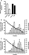
Effects of PKA modulators on ApoB and ApoE secretion and HCV particle buoyant density. (A) Extracellular medium was collected from control and H89 (10 μM)-treated Huh-7.5 cells, and the levels of ApoB (white bars) and ApoE (black bars) were measured by capture ELISA. Data are expressed relative to those for control untreated cells and represent the mean of data for three replicate infections. The buoyant density (•) of extracellular J6/JFH released from control (B) or H89-treated (C) Huh-7.5 cells was determined on iodixanol gradients. Individual bars show relative HCV RNA copy numbers in each fraction compared to the maximum peak.
FIG. 10.
FIG. 10.
HCV infection increases cAMP levels and PKA activity. (A) cAMP levels were measured in uninfected and J6/JFH- and JFH-1-infected Huh-7.5 cells 72 h postinfection. As a control, Huh-7.5 cells were incubated with FK (10 μM), a compound known to activate adenylyl cyclase and increase cAMP levels. cAMP levels are shown relative to control untreated cells and represent data from the means of three replicate wells. (B) PKA activity was assessed by measuring the reactivity of an anti-PKA substrate-specific antibody (p-PKAs) with 10 μg of total protein separated by SDS-PAGE from control (lane 1), FK (10 μM)-stimulated (lane 2), and JFH-1-infected (72 h postinfection) (lane 3) Huh-7.5 cells. (C) CLDN1 and PKA substrates were immunoprecipitated from 100 μg of uninfected (UF) and JFH-1-infected (72 h postinfection) Huh-7.5 cell lysates with specific antibodies (mouse anti-CLDN1, rabbit anti-CLDN1, and p-PKAs) and control antibodies (murine IgG [mIgG] and rabbit Ig [rIgG]). Immunoprecipitates were subjected to SDS-PAGE, and reactivity with rabbit anti-CLDN1 was assessed by Western blotting. (D and E) Uninfected (D) and JFH-1-infected (E) (72 h postinfection) Huh-7.5 cells were incubated with FK or H89 for 1 h (H89 > FK indicates a 1-h preincubation with H89 prior to FK treatment), fixed, and stained with the PKA substrate-specific antibody (p-PKAs) (green). JFH-1-infected cells were visualized by staining for NS5A (red), and nuclei were counterstained with DAPI (blue). Laser scanning confocal microscopic images of single 1-μm z sections were obtained using a 63× 1.2-numerical-aperture objective (scale bar represents 10 μm).

Similar articles

Cited by

References

    1. Alto, N. M., S. H. Soderling, N. Hoshi, L. K. Langeberg, R. Fayos, P. A. Jennings, and J. D. Scott. 2003. Bioinformatic design of A-kinase anchoring protein-in silico: a potent and selective peptide antagonist of type II protein kinase A anchoring. Proc. Natl. Acad. Sci. USA 1004445-4450. - PMC - PubMed
    1. Amella, C. A., B. Sherry, D. H. Shepp, and H. Schmidtmayerova. 2005. Macrophage inflammatory protein 1α inhibits postentry steps of human immunodeficiency virus type 1 infection via suppression of intracellular cyclic AMP. J. Virol. 795625-5631. - PMC - PubMed
    1. Andrade, A. A., P. N. Silva, A. C. Pereira, L. P. De Sousa, P. C. Ferreira, R. T. Gazzinelli, E. G. Kroon, C. Ropert, and C. A. Bonjardim. 2004. The vaccinia virus-stimulated mitogen-activated protein kinase (MAPK) pathway is required for virus multiplication. Biochem. J. 381437-446. - PMC - PubMed
    1. Aoubala, M., J. Holt, R. A. Clegg, D. J. Rowlands, and M. Harris. 2001. The inhibition of cAMP-dependent protein kinase by full-length hepatitis C virus NS3/4A complex is due to ATP hydrolysis. J. Gen. Virol. 821637-1646. - PubMed
    1. Avila-Flores, A., E. Rendon-Huerta, J. Moreno, S. Islas, A. Betanzos, M. Robles-Flores, and L. Gonzalez-Mariscal. 2001. Tight-junction protein zonula occludens 2 is a target of phosphorylation by protein kinase C. Biochem. J. 360295-304. - PMC - PubMed

Publication types

MeSH terms

LinkOut - more resources