. 2008 Sep;82(17):8383-91.
doi: 10.1128/JVI.00348-08. Epub 2008 Jun 25.

Changes in p19Arf localization accompany apoptotic crisis during pre-B-cell transformation by Abelson murine leukemia virus

Affiliations

Changes in p19Arf localization accompany apoptotic crisis during pre-B-cell transformation by Abelson murine leukemia virus

Rebekah Stackpole Zimmerman et al. J Virol. 2008 Sep.

Abstract

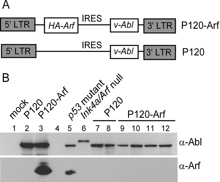
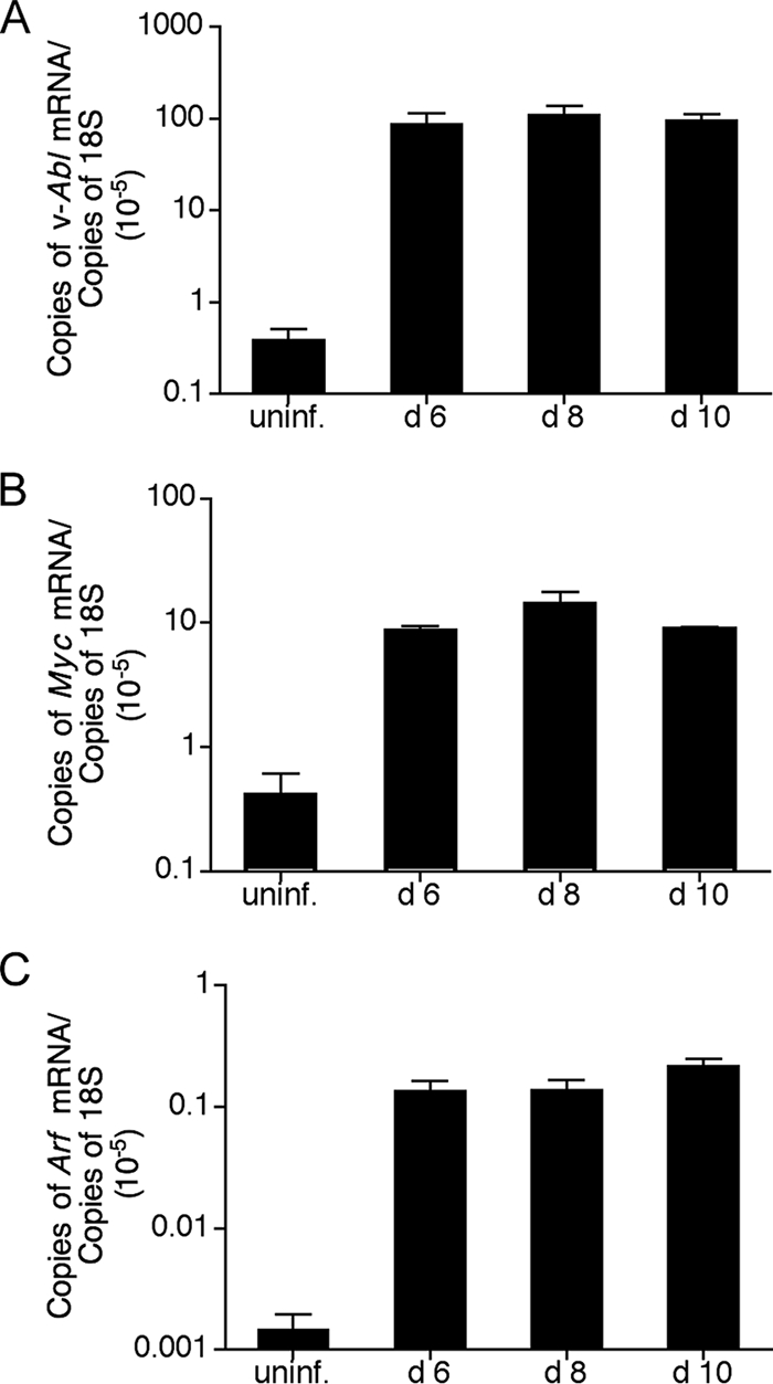
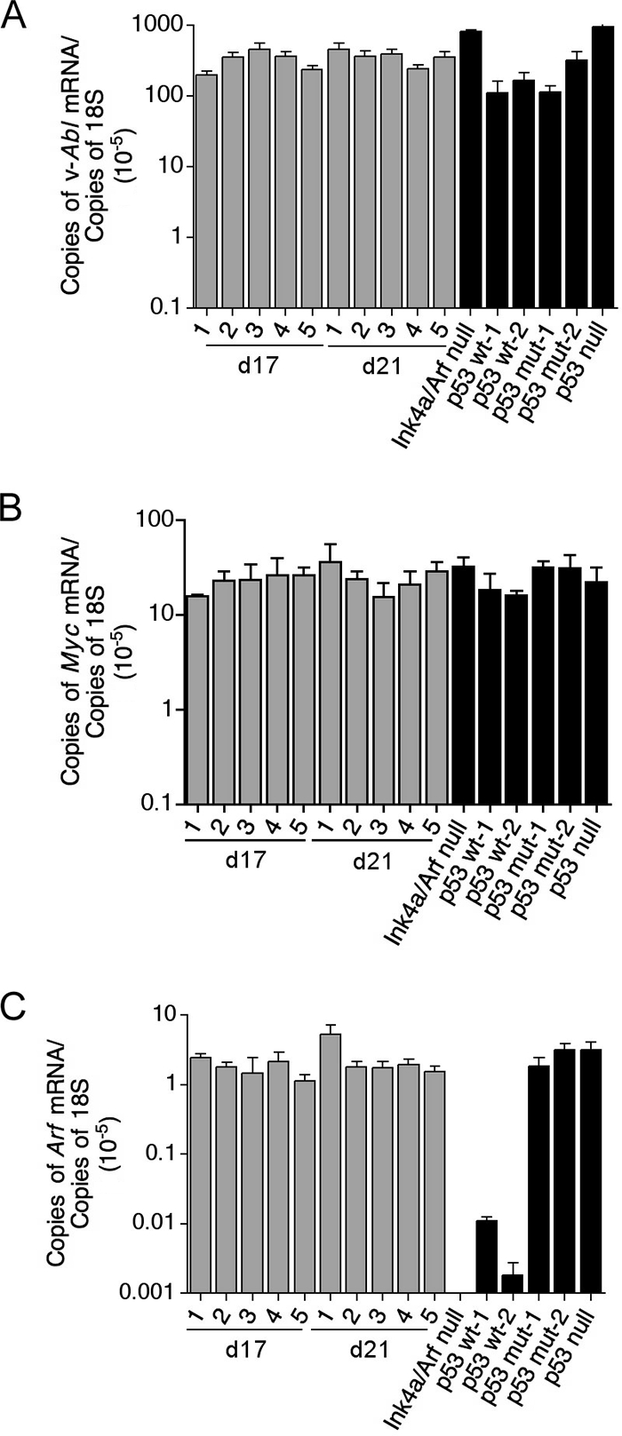
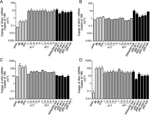
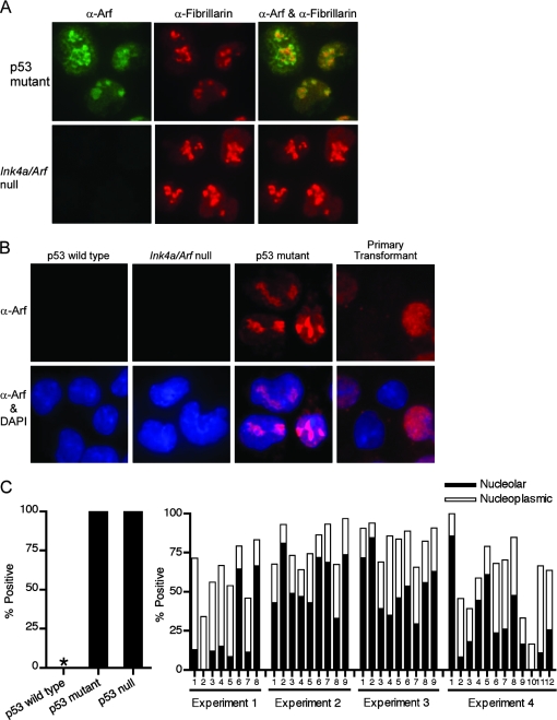
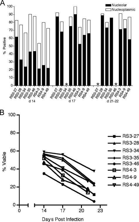
Transformation by Abelson murine leukemia virus (Ab-MLV) is a multistep process in which growth-stimulatory signals from the v-Abl oncoprotein and growth-suppressive signals from the p19(Arf)-p53 tumor suppressor pathway oppose each other and influence the outcome of infection. The process involves a proliferative phase during which highly viable primary transformants expand, followed by a period of marked apoptosis (called "crisis") that is dependent on the presence of p19(Arf) and p53; rare cells that survive this phase emerge as fully transformed and malignant. To understand the way in which v-Abl expression affects p19(Arf) expression, we examined changes in expression of Arf during all stages of Ab-MLV transformation process. As is consistent with the ability of v-Abl to stimulate Myc, a transcription factor known to induce p19(Arf), Myc and Arf are induced soon after infection and p19(Arf) is expressed. At these early time points, the infected cells remain highly viable. The onset of crisis is marked by an increase in p19(Arf) expression and a change in localization of the protein from the nucleoplasm to the nucleolus. These data together suggest that the localization and expression levels of p19(Arf) modulate the effects of the protein during oncogenesis and reveal that the p19(Arf)-mediated response is subject to multiple layers of regulation that influence its function during Ab-MLV-mediated transformation.


Figures

FIG. 1.
FIG. 1.
Expression of p19Arf can inhibit transformation in pre-B cells. (A) A schematic diagram of the P120-Arf and P120 viruses. IRES, internal ribosome entry site; LTR, long terminal repeat. (B) Lanes 1 to 3, 293T cells were transfected with 15 μg of viral DNA encoding either P120-Arf or P120 or were subjected to mock transfection; lysates were prepared 48 h posttransfection. Lanes 5 to 12, lysates were prepared from either control cell lines (p53 mutant and Ink4a/Arf null) or cell lines transformed with the Ab-MLV P120 strain, the Ab-MLV P160 strain (lane 6), or the P120-Arf virus and lysates were analyzed using Western blotting with antibodies against Gag/v-Abl (3) and p19Arf.
FIG. 2.
FIG. 2.
v-abl, Myc, and Arf are induced early in transformation. RNA was harvested from uninfected B220+ CD43+ cells (uninf.) and infected cells 6, 8, and 10 days after infection with Ab-MLV and reverse transcribed with random hexamer primers. Real-time PCR using primers specific for v-abl (A), Myc (B), and Arf (C) was performed, and results were normalized to 18S RNA results. Error bars represent the standard deviations of the means of the results of three independent PCRs from one reverse transcription. The data are representative of values obtained for at least two independent infections in which cDNAs from two independent reverse transcriptions were analyzed in triplicate.
FIG. 3.
FIG. 3.
Arf mRNA expression increases as cells enter crisis. RNA was harvested from five different infected cultures 17 and 21 days after infection with Ab-MLV (gray bars) as well as from cultures of six different established cell lines of the indicated genotypes (black bars). The RNA was reverse transcribed with random hexamer primers, followed by real-time PCR using primers specific for v-abl (A), Myc (B), and Arf (C). Results were normalized to 18S RNA results. Error bars represent the standard deviations of the means of the results of three PCRs from one reverse transcription. The data are representative of values obtained for at least two independent infections in which cDNAs from two independent reverse transcriptions were analyzed in triplicate.
FIG. 4.
FIG. 4.
Genes involved in the Arf-p53 pathway show subtle changes in expression during transformation. Real-time PCR was performed using the same cDNAs described for Fig. 2 and 3 with primers specific for Dmp1 (A), Bmi-1 (B), p53 (C), and Bax (D), and the results were normalized to 18S RNA results. Error bars represent the standard deviations of the means of the results of three PCRs from one reverse transcription. The data are representative of values obtained for at least two independent infections in which cDNAs from two independent reverse transcriptions were analyzed in triplicate. uninf., uninfected.
FIG. 5.
FIG. 5.
Some primary transformants do not express nucleolar p19Arf. (A) Control cell lines expressing mutant p53 or derived from Ink4a/Arf null mice were costained with antibodies against p19Arf and fibrillarin, a nucleolar marker (27). (B) Primary transformants and control cell lines were stained with antibodies directed against p19Arf and with DAPI and examined using a fluorescence microscope. The images shown are representative of analysis of at least 100 cells in each preparation. Primary transformants were examined 10 days postinfection. (C) The frequency of cells expressing p19Arf in the nucleolus or in the nucleoplasm was determined by examining the staining pattern in control cells (left panel) and in primary transformants (right panel). A minimum of 100 cells were scored for each sample. Examples of results obtained with primary transformants examined 10 days postinfection and derived from four independent experiments are shown. Black bars indicate nucleolar localization; white bars indicate localization in the nucleoplasmic region.
FIG. 6.
FIG. 6.
Ab-MLV transformants express NPM in the cytoplasm. (A) NPM expression was examined by Western blotting using uninfected B220+ CD43+ pre-B cells (Uninfected), primary transformants examined 10 days postinfection, and a panel of fully transformed cell lines of the indicated genotypes. The blot was stripped and reprobed with antibodies against β-actin to control for loading. (B) Whole bone marrow, uninfected B220+ CD43+ cells, primary transformants, and cells in the crisis phase of transformation were stained with antibodies against NPM and with DAPI and examined by immunofluorescence microscopy. A minimum of 100 cells were examined for each sample.
FIG. 7.
FIG. 7.
The percent of cells with nucleolar p19Arf increases as cells enter crisis. Primary transformants were plated in liquid medium and analyzed during the outgrowth and crisis period. (A) Cells were stained with antibodies directed against p19Arf 14, 17, 21, and 22 days after infection and examined by immunofluorescence microscopy for the frequency of cells expressing p19Arf in the nucleolus and in the nucleoplasm. At least 100 cells were examined for each sample. The black bars indicate nucleolar localization; the white bars indicate nucleoplasmic localization; an asterisk indicates that too few cells were available for analysis. (B) The viability of cells in the cultures examined as described for panel A was determined by counting trypan blue-stained cells with a hemocytometer.

Similar articles

Cited by

References

    1. Apicelli, A. J., L. B. Maggi, Jr., A. C. Hirbe, A. P. Miceli, M. E. Olanich, C. L. Schulte-Winkeler, A. J. Saporita, M. Kuchenreuther, J. Sanchez, K. Weilbaecher, and J. D. Weber. 2008. A non-tumor suppressor role for basal p19ARF in maintaining nucleolar structure and function. Mol. Cell. Biol. 281068-1080. - PMC - PubMed
    1. Baughn, L. B., and N. Rosenberg. 2005. Disruption of the Shc/Grb2 complex during Abelson virus transformation affects proliferation, but not apoptosis. J. Virol. 792325-2334. - PMC - PubMed
    1. Chesebro, B., K. Wehrly, M. Cloyd, W. Britt, J. Portis, J. Collins, and J. Nishio. 1981. Characterization of mouse monoclonal antibodies specific for Friend murine leukemia virus-induced erythroleukemia cells: Friend-specific and FMR-specific antigens. Virology 112131-144. - PubMed
    1. Colombo, E., P. Bonetti, E. Lazzerini Denchi, P. Martinelli, R. Zamponi, J. C. Marine, K. Helin, B. Falini, and P. G. Pelicci. 2005. Nucleophosmin is required for DNA integrity and p19Arf protein stability. Mol. Cell. Biol. 258874-8886. - PMC - PubMed
    1. Falini, B., C. Mecucci, E. Tiacci, M. Alcalay, R. Rosati, L. Pasqualucci, R. La Starza, D. Diverio, E. Colombo, A. Santucci, B. Bigerna, R. Pacini, A. Pucciarini, A. Liso, M. Vignetti, P. Fazi, N. Meani, V. Pettirossi, G. Saglio, F. Mandelli, F. Lo-Coco, P. G. Pelicci, and M. F. Martelli. 2005. Cytoplasmic nucleophosmin in acute myelogenous leukemia with a normal karyotype. N. Engl. J. Med. 352254-266. - PubMed

Publication types

MeSH terms