. 2008 Oct;19(10):1919-28.
doi: 10.1681/ASN.2007111216. Epub 2008 Jun 25.

Activator protein 2alpha mediates parathyroid TGF-alpha self-induction in secondary hyperparathyroidism

Affiliations

Activator protein 2alpha mediates parathyroid TGF-alpha self-induction in secondary hyperparathyroidism

Maria Vittoria Arcidiacono et al. J Am Soc Nephrol. 2008 Oct.

Abstract

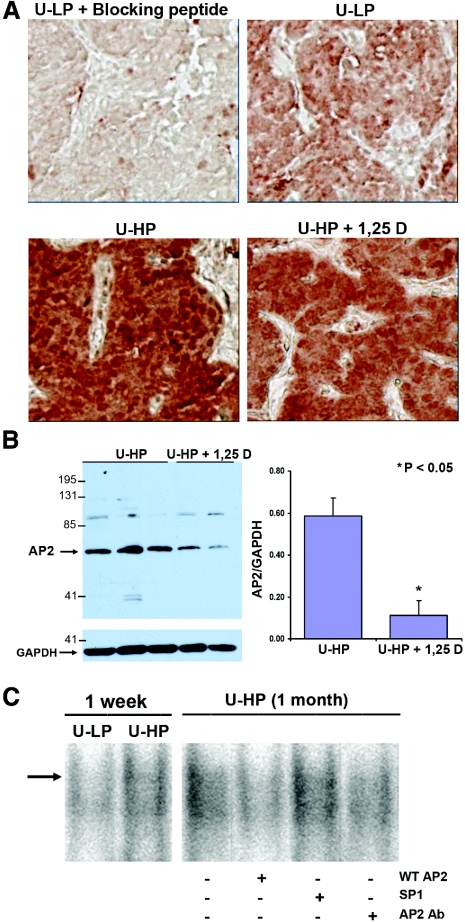
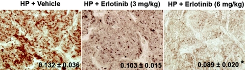
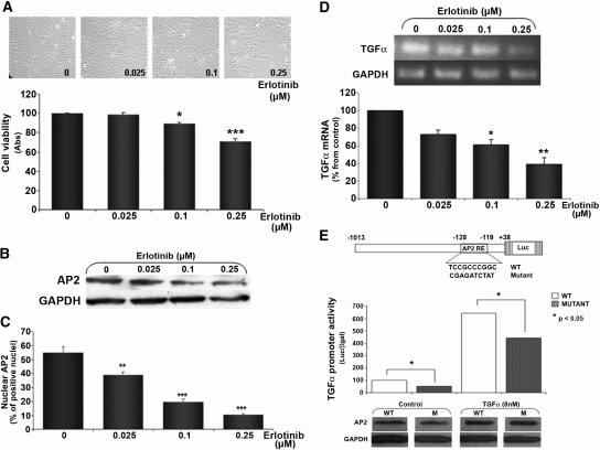
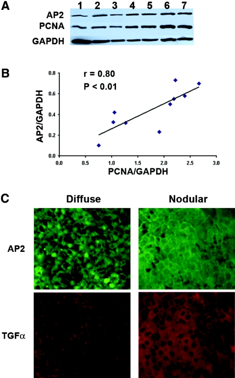
In secondary hyperparathyroidism, enhanced expression of TGF-alpha in the parathyroid leads to its own upregulation, generating a feed-forward loop for TGF-alpha activation of its receptor, EGFR receptor (EGFR), which promotes parathyroid hyperplasia. These studies examined the role of activator protein 2alpha (AP2), an inducer of TGF-alpha gene transcription, in the upregulation of parathyroid TGF-alpha in secondary hyperparathyroidism. In rat and human secondary hyperparathyroidism, parathyroid AP2 expression strongly correlated with TGF-alpha levels and with the rate of parathyroid growth, as expected. Furthermore, the increases in rat parathyroid content of AP2 and its binding to a consensus AP2 DNA sequence preceded the increase in TGF-alpha induced by high dietary phosphate. More significant, in A431 cells, which provide a model of enhanced TGF-alpha and TGF-alpha self-induction, mutating the core AP2 site of the human TGF-alpha promoter markedly impaired promoter activity induced by endogenous or exogenous TGF-alpha. Important for therapy, in five-sixths nephrectomized rats fed high-phosphate diets, inhibition of parathyroid TGF-alpha self-induction using erlotinib, a highly specific inhibitor of TGF-alpha/EGFR-driven signals, reduced AP2 expression dosage dependently. This suggests that the increases in parathyroid AP2 occur downstream of EGFR activation by TGF-alpha and are required for TGF-alpha self-induction. Indeed, in A431 cells, erlotinib inhibition of TGF-alpha self-induction caused parallel reductions in AP2 expression and nuclear localization, as well as TGF-alpha mRNA and protein levels. In summary, increased AP2 expression and transcriptional activity at the TGF-alpha promoter determine the severity of the hyperplasia driven by parathyroid TGF-alpha self-upregulation in secondary hyperparathyroidism.


Figures

Figure 1.
Figure 1.
(A) Parathyroid AP2 immunostaining in response to changes in dietary P intake and/or prophylactic calcitriol administration in rat SH. Representative photomicrographs of immunohistochemical staining of AP2 in parathyroid tissue from five-sixths Nx rats undergoing the experimental conditions described in protocol 1, at day 7 after the onset of renal failure: Uremic (U) + high-P diet (U-HP); U + low-P (LP) diet (U-LP); U-HP diet + 1,25D (U-HP + 1,25D). (B) Parathyroid AP2 expression in response to prophylactic calcitriol administration in rat SH. Western blot analysis and densitometric quantification of parathyroid AP2/GAPDH ratios in parathyroid glands from five-sixths Nx rats fed high dietary P and receiving either vehicle or calcitriol (1,25D). Bars and error bars represent means ± SEM; *P < 0.05 (see Statistical Analysis section). (C) Dietary P regulation of parathyroid AP2/DNA binding. Binding to a radiolabeled AP2 consensus sequence of nuclear extracts (30 μg of total protein) from parathyroid glands from five-sixths Nx (U) rats fed either HP (0.9% P; U-HP) or LP (0.2% P; U-LP) for 1 wk. The specificity of AP2/DNA binding was studied with similar incubations of radiolabeled AP2 consensus with nuclear extracts from hyperplastic parathyroid glands from uremic rats fed high dietary P for 1 mo (U-HP [1 mo]) in the absence or presence of a 200-mol excess of radioinert AP2 consensus oligonucleotide (WT AP2) or SP1, an AP2-unrelated oligonucleotide sequence (SP1), or 1 μl of a polyclonal antibody against AP2α (AP2 Ab). Magnification, ×400 in A.
Figure 2.
Figure 2.
Erlotinib inhibits the enhancement of parathyroid AP2 induced by kidney disease and high dietary P. Representative photomicrographs of immunohistochemical staining of AP2 in parathyroid tissue at day 7 after five-sixths Nx in rats fed HP (1.2%) receiving vehicle (HP+Vehicle) or 3 or 6 mg erlotinib/kg body wt (HP+Erlotinib 3 or 6 mg/kg, respectively). Values indicate the results of the immunohistochemical quantification of the changes in parathyroid AP2 content in response to erlotinib administration in rats treated as described. Values represent means ± SEM of parathyroid AP2 measurements from six rats; *P < 0.05 versus HP+Vehicle. Magnification, ×200.
Figure 3.
Figure 3.
AP2 contribution to TGF-α self-induction in A431 cells. (A) Erlotinib dosage-dependent inhibition of TGF-α/EGFR–driven growth as measured by progressive reduction in cell number (top) and MTT assay (bottom). Bars and error bars indicate means ± SEM of erlotinib-induced reduction in viable cells compared with untreated controls (untreated, erlotinib 0 μM) from four independent experiments. (B) Western blot analysis of changes in AP2 expression in response to increasing dosages of erlotinib in A431 cells treated as in A. (C) Quantification of changes in nuclear AP2 expression using immunofluorescent staining for AP2, in response to erlotinib. Bars and error bars represent means ± SEM of the number of AP2-positive nuclei per 1000 cells from three independent experiments. Results are expressed as percentage of nuclear AP2 content relative to untreated (erlotinib 0 μM) cells. (D) Erlotinib dosage-dependent inhibition of TGF-α mRNA levels in cells treated as in A. (Top) Representative RT-PCR of TGF-α mRNA expression in response to erlotinib. (Bottom) Bars and error bars represent the means ± SEM of TGF-α mRNA levels, corrected per GAPDH, from two independent experiments. Results are expressed as percentage of untreated A431 cells (0). (E, top) Partial sequence of the human TGF-α promoter containing an AP2 binding site (wild-type) and the insertion of a 10-bp mutation at the core AP2 consensus (mutant). (E, middle) Promoter activity in response to TGF-α treatment in A431 cells transfected with wild-type (WT; □) or mutant (□) TGF-α promoter/luciferase reporter. Bars and error bars indicate means ± SEM from triplicate luciferase/β-gal determinations per experimental condition from two independent experiments (P < 0.05). (E, bottom) Representative Western blot analysis of TGF-α (8 nM) induction of AP2 levels after 24 h of treatment in the cell lysates from A431 cells transfected as indicated for luciferase reporter assays.
Figure 4.
Figure 4.
(A) Parathyroid AP2 expression correlates directly with PCNA in human SH. (Top) Western blot analysis of AP2, PCNA, and GAPDH content in whole-cell extracts from 10 human parathyroid glands with no pathologic diagnosis of diffuse or nodular hyperplasia. (Bottom) Correlation between AP2 and PCNA content, as measured by densitometric analysis of AP2/GAPDH and PCNA/GAPDH ratios in whole-cell extracts from 10 hyperplastic human parathyroid glands depicted in the Western blot. (B) Parathyroid AP2 expression correlates directly with TGF-α content in human SH. Representative photomicrographs of immunofluorescence staining for AP2 and TGF-α in diffuse and nodular human parathyroid glands. Magnification, ×200.
Figure 5.
Figure 5.
Model for AP2 regulation of TGF-α self-induction. The release of the mature form of TGF-α from its cell membrane precursor binds and activates its receptor, the EGFR, thereby inducing increases in AP2 expression, nuclear translocation, and transactivation of the TGF-α gene. Inhibition of EGFR activation with erlotinib is sufficient to simultaneously arrest increases in AP2 and AP2-mediated TGF-α gene transcription.

References

    1. Goodman WG, Goldin J, Kuizon BD, Yoon C, Gales B, Sider D, Wang Y, Chung J, Emerick A, Greaser L, Elashoff RM, Salusky IB: Coronary-artery calcification in young adults with end-stage renal disease who are undergoing dialysis. N Engl J Med 342: 1478–1483, 2000 - PubMed
    1. Cozzolino M, Dusso AS, Slatopolsky E: Role of calcium-phosphate product and bone-associated proteins on vascular calcification in renal failure. J Am Soc Nephrol 12: 2511–2516, 2001 - PubMed
    1. Gonzalez EA, Martin KJ: Renal osteodystrophy: Pathogenesis and management. Nephrol Dial Transplant 10[Suppl 3]: 13–21, 1995 - PubMed
    1. Parfitt AM: The hyperparathyroidism of chronic renal failure: A disorder of growth. Kidney Int 52: 3–9, 1997 - PubMed
    1. Slatopolsky E, Finch J, Denda M, Ritter C, Zhong M, Dusso A, MacDonald PN, Brown AJ: Phosphorus restriction prevents parathyroid gland growth: High phosphorus directly stimulates PTH secretion in vitro. J Clin Invest 97: 2534–2540, 1996 - PMC - PubMed

Publication types

MeSH terms

Substances