. 2008 Jun 25;28(26):6670-8.
doi: 10.1523/JNEUROSCI.1702-08.2008.

NMDA receptor blockade with memantine attenuates white matter injury in a rat model of periventricular leukomalacia

Affiliations

NMDA receptor blockade with memantine attenuates white matter injury in a rat model of periventricular leukomalacia

Simon M Manning et al. J Neurosci. .

Abstract

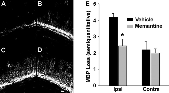
Hypoxia-ischemia (H/I) in the premature infant leads to white matter injury termed periventricular leukomalacia (PVL), the leading cause of subsequent neurological deficits. Glutamatergic excitotoxicity in white matter oligodendrocytes (OLs) mediated by cell surface glutamate receptors (GluRs) of the AMPA subtype has been demonstrated as one factor in this injury. Recently, it has been shown that rodent OLs also express functional NMDA GluRs (NMDARs), and overactivation of these receptors can mediate excitotoxic OL injury. Here we show that preterm human developing OLs express NMDARs during the PVL period of susceptibility, presenting a potential therapeutic target. The expression pattern mirrors that seen in the immature rat. Furthermore, the uncompetitive NMDAR antagonist memantine attenuates NMDA-evoked currents in developing OLs in situ in cerebral white matter of immature rats. Using an H/I rat model of white matter injury, we show in vivo that post-H/I treatment with memantine attenuates acute loss of the developing OL cell surface marker O1 and the mature OL marker MBP (myelin basic protein), and also prevents the long-term reduction in cerebral mantle thickness seen at postnatal day 21 in this model. These protective doses of memantine do not affect normal myelination or cortical growth. Together, these data suggest that NMDAR blockade with memantine may provide an effective pharmacological prevention of PVL in the premature infant.


Figures

Figure 1.
Figure 1.
NR1, the NMDAR obligate subunit, is present on multiple white matter cell types in the P6 Long–Evans rat pup during the window of susceptibility for PVL. Double-fluorescent staining of normal P6 rat pup 50 μm frozen sections is shown. A, D, G, J, Cell-specific antibody labeling of developing OLs (O4), astrocytes (GFAP), microglia (CD11), and subplate neurons (NeuN). B, E, H, K, NR1 obligate NMDAR antibody labeling and nuclear DAPI stain. C, F, I, L, Overlay showing representative NR1 expression pattern within these four white matter cell types. M, Confocal Z-stack of O4 (red), NR1 (green), and DAPI (blue), showing colocalization of NR1 with O4-positive developing OL processes. Scale bars: C, F, I, 10 μm; L, M, 50 μm.
Figure 2.
Figure 2.
Preterm human periventricular white matter developing OLs express NR1, the NMDAR obligate subunit, during the window of susceptibility for PVL. A–F, Human O4-positive (late progenitor and immature premyelinating) developing OLs express NR1, predominantly in their processes, at both 26 (A–C, arrowheads) and 31 (D–F) PCW. Other O4-negative white matter cell types also express NR1 (E, F, arrows). Scale bars, 20 μm. G, Confocal Z-stack of the immature premyelinating OL marker, O1 (red), and NR1 (green), showing colocalization of NR1 with O1 throughout premyelinating OL processes. Scale bars, 20 μm.
Figure 3.
Figure 3.
Memantine significantly attenuates loss of the developing OL cell surface marker O1 ipsilateral to the carotid ligation at 72 h after injury. A–D, Representative O1 immunohistochemistry showing loss of O1 staining, more severe ipsilateral (A, C) to the carotid artery ligation, in vehicle-treated pups (A, B), with attenuation of O1 loss in memantine-treated pups (C, D). Scale bar, 100 μm. E, Ipsilateral O1 loss represented as a ratio of contralateral stain (1 = no loss; 0 = complete loss). Memantine significantly attenuates loss of the O1 marker of developing OLs (n = 11 vehicle; n = 12 memantine; t test, p = 0.012). Error bars denote SEM.
Figure 4.
Figure 4.
Memantine significantly attenuates loss of MBP at 72 h after injury. A–D, Representative MBP immunohistochemistry: vehicle-treated (A, B) and memantine-treated (C, D), ipsilateral (A, C) and contralateral (B, D) to carotid artery ligation. Scale bar, 100 μm. E, MBP loss based on a five-point semiquantitative scale (0 = no loss; 5 = maximum loss). Memantine significantly attenuates MBP loss ipsilateral to carotid artery ligation (n = 11 vehicle; n = 14 memantine; t test, p = 0.008). Error bars denote SEM.
Figure 5.
Figure 5.
Memantine reduces long-term sequelae of white matter injury at 15 d after injury. ImageJ quantification of cerebral mantle (CM) thickness represented as ratio of ipsilateral (to carotid ligation) to contralateral values (1 = no loss; 0 = complete loss) (n = 9 vehicle; n = 10 memantine; Mann–Whitney rank sum test, p = 0.037). Error bars denote SEM.
Figure 6.
Figure 6.
NMDA-evoked currents in P6–P7 rat white matter OLs in situ are significantly attenuated by memantine in a dose-dependent manner. A, B, Raw data showing NMDA-evoked current in developing OL after application of 50 μm NMDA and attenuation with application of 10 μm (A) and 50 μm (B) memantine. C, Bar graph showing percentage reduction in NMDA peak current amplitudes at negative and positive voltages after application of 10 μm memantine (n = 5 recordings) and 50 μm memantine (n = 4 recordings) normalized to control peak currents (n = 6 recordings). Memantine significantly reduced peak current amplitudes at −15 mV (1-way ANOVA, p = 0.036) and at +50 mV (1-way ANOVA, p = 0.024). Error bars denote SEM. D, E, We identified cells as developing OLs by current signature, morphology, and immunohistochemistry. D, The outward and inward currents in morphologically identified developing OLs in P6 white matter slice are typical of developing OLs. Currents were recorded by stepping cells from −90 mV to +50 mV in 10 mV intervals. The calibration is indicated. E, Example of confocal projection of a recorded cell injected with the fluorescent dye Alexa Fluor 594 (red) and stained for the nuclear pan-OL marker Olig2 (green), confirming that memantine attenuation of NMDA-evoked currents occurs in OLs. Scale bar, 10 μm.

References

    1. Back SA, Luo NL, Borenstein NS, Levine JM, Volpe JJ, Kinney HC. Late oligodendrocyte progenitors coincide with the developmental window of vulnerability for human perinatal white matter injury. J Neurosci. 2001;21:1302–1312. - PMC - PubMed
    1. Bakiri Y, Hamilton NB, Káradóttir R, Attwell D. Testing NMDA receptor block as a therapeutic strategy for reducing ischaemic damage to CNS white matter. Glia. 2008;56:233–240. - PMC - PubMed
    1. Block F, Schwarz M. Memantine reduces functional and morphological consequences induced by global ischemia in rats. Neurosci Lett. 1996;208:41–44. - PubMed
    1. Cai Z, Lin S, Fan LW, Pang Y, Rhodes PG. Minocycline alleviates hypoxic-ischemic injury to developing oligodendrocytes in the neonatal rat brain. Neuroscience. 2006;137:425–435. - PubMed
    1. Chen HS, Lipton SA. The chemical biology of clinically tolerated NMDA receptor antagonists. J Neurochem. 2006;97:1611–1626. - PubMed

Publication types

MeSH terms

LinkOut - more resources