. 2008 Jun 25;28(26):6742-9.
doi: 10.1523/JNEUROSCI.1082-08.2008.

Calcium control of endocytic capacity at a CNS synapse

Affiliations

Calcium control of endocytic capacity at a CNS synapse

J Balaji et al. J Neurosci. .

Abstract

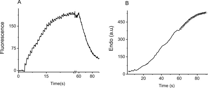
The ability to recycle synaptic vesicles is a crucial property of nerve terminals that allows maintenance of synaptic transmission. Using high-sensitivity optical approaches at hippocampal nerve terminals in dissociated neurons in culture, we show that modulation of endocytosis can be achieved by expansion of the endocytic capacity. Our experiments indicate that the endocytic capacity, the maximum number of synaptic vesicles that can be internalized in parallel at individual synapses, is tightly controlled by intracellular calcium levels. Increasing levels of intracellular calcium, which occurs as firing frequency increases, significantly increases the endocytic capacity. At physiological temperature after 30 Hz firing, these synapses are capable of endocytosing at least approximately 28 vesicles in parallel, each with a time constant of approximately 6 s. This calcium-dependent control of endocytic capacity reveals a potentially useful adaptive response to high-frequency activity to increase endocytic rates under conditions of vesicle pool depletion.


Figures

Figure 1.
Figure 1.
A, B, The rate of decay of synaptopHluorin (A) and vGlut-pHluorin (B) fluorescence after action potential stimulation reaches a limiting value during a prolonged stimulus. The lines in A and B are the fluorescence response of synaptopHluorin- and vGpH-transfected synaptic boutons, respectively, that are subject to varying duration of stimulus (50, 70, 100, 150, 200, 300, 400, and 500 AP at 10 Hz for spH; 50, 75, 100, 150, 200, 250, and 300 at 10 Hz AP for vGpH). The peak of the pHluorin response is proportional to surface accumulation of the vesicular membrane at the end of the stimulus. The dashed curves are the response of the same set of boutons for action potentials fired at 33 Hz for 10 s, which provides a measure of the total recycling pool.
Figure 2.
Figure 2.
Poststimulus decay in pHluorin response fits well to single-exponential decay. A, B, Open circles are the average fluorescent traces of boutons transfected with spH and vGpH, respectively (∼30 boutons in each case). The line is the exponential fit to the data. C, D, The time constants for synaptopHluorin- (n = 3 cells, 45–60 boutons each) and vGpH- (n = 4 cells, 30–50 boutons each) transfected neurons measured as a function of surface accumulation. The decay constants start to increase after ∼45% for each pHluorin-tagged synaptic vesicle protein. The increasing decay constants fit to a straight line. The point at which this line intersects the fundamental time constant (normalized to unity) yields the Cendo of the cell. For spH (C), the mean time constant in the nonsaturating region is 19.5 ± 1.3 s, and for vGpH (D), it is 14.2 ± 2.2 s.
Figure 3.
Figure 3.
Exocytosis and endocytosis during persistent stimulation followed in real time. A, Responses of three boutons for single action potentials delivered every 15 s (arrows). B, The ensemble average time trace obtained for stimuli delivered at 2, 5, 7, 10, 12, and 15 s intervals from the same 39 boutons in each run. C, Cumulative exocytosis (circles), surface and alkaline pool (squares), and apparent cumulative endocytosis (triangles) calculated from 2 s interval run shown in B as described in the Appendix. D, Endocytosis curves for the 2, 5, and 15 s stimulus interval (squares, circles, and triangles, respectively), calculated as in C, are fit linearly in the time region in which the surface fluorescence is at a quasi-steady state to obtain the endocytic rates in this regime. E, The time constants for endocytosis derived from the endocytic rates and surface accumulations (as described in the Appendix) for different stimulus intervals are shown (5 cells, 372 boutons). a.f.u., Arbitrary fluorescence units; a.u., arbitrary units.
Figure 4.
Figure 4.
Endocytosis rate during and after stimulation is very similar. A, The vGpH fluorescence curve during stimulation at 0.5 Hz is compared with the vGpH fluorescence trace obtained after the stimulus for the same set of synapses. B, The derived cumulative endocytosis curve (black) during stimulation obtained from subtracting the surface accumulation from the cumulative exocytosis curve is very similar to the vGpH fluorescence decay (open symbols, here inverted for comparison), which is dominated by endocytosis after the stimulus. a.u., Arbitrary units.
Figure 5.
Figure 5.
Endocytic capacity of the neuron is modulated by altering the levels of intracellular calcium. A, B, Plots of decay constants obtained for synaptopHluorin (5 cells) and vGpH (4 cells) obtained using varying stimulus duration (6–50 s) at 10 Hz (black circles) and 33 Hz (gray squares). A, For spH, the average time constant in the nonsaturating region is 19.5 ± 1.3 s for the 10 Hz and 19.6 ± 1.4 s for the 33 Hz data. B, For vGpH, the average time constant in the nonsaturating region is 14.2 ± 2.2 s for the 10 Hz and 15.0 ± 1.9 s for the 33 Hz data. Endocytosis saturates at higher accumulation, suggesting an increased endocytic capacity at higher frequency of stimulation. The “endocytic capacity” defined by abscissa of where the straight line fit (dashed line in both graphs) is unity.
Figure 6.
Figure 6.
Endocytic capacity measured as a function of relative intracellular calcium. Fluorescent signal of calcium indicators (Mag Green) is used as a relative measure of intracellular steady-state calcium during the stimulus. A, The fluorescence measured as a function of time for 10 (light gray), 20 (dark gray), and 30 (black) Hz stimulus at 2 mm external calcium measured at an individual synaptic varicosity. B, Endocytic capacity, Cendo (circles), is plotted against the relative intracellular steady-state calcium levels obtained by changing either the stimulus frequency while keeping external CaCl2 at 2 mm (filled symbols, 10, 20, and 30 Hz) or external calcium concentration while keeping the stimulus frequency at 10 Hz (open symbols, 1, 2, and 3 mm CaCl2) (8 cells).
Figure 7.
Figure 7.
Fundamental time constant is faster at 36°C. A, Comparison of the fluorescence signal after a 150 AP stimulus for the same set of 49 boutons at 28°C (black), which yields τ = 14.96 ± 0.08, and at 36°C (gray), yielding τ = 5.79 ± 0.04. B, Saturation of the endocytic capacity is apparent at 36°C during 30 Hz stimulation. Synapses were stimulated with varying numbers of AP (15–450) at 30 Hz, and the time constants for vGpH decays were obtained for each accumulation level. These data were obtained from 10 cells (31–93 boutons each), and the time constants for a given cell were normalized to the smallest value in the nonsaturating region of each cell. The average time constant in the nonsaturating region is 6 ± 1.3 s.

Similar articles

Cited by

References

    1. Anggono V, Smillie KJ, Graham ME, Valova VA, Cousin MA, Robinson PJ. Syndapin I is the phosphorylation-regulated dynamin I partner in synaptic vesicle endocytosis. Nat Neurosci. 2006;9:752–760. - PMC - PubMed
    1. Atluri PP, Regehr WG. Determinants of the time course of facilitation at the granule cell to Purkinje cell synapse. J Neurosci. 1996;16:5661–5671. - PMC - PubMed
    1. Atluri PP, Ryan TA. The kinetics of synaptic vesicle reacidification at hippocampal nerve terminals. J Neurosci. 2006;26:2313–2320. - PMC - PubMed
    1. Balaji J, Ryan TA. Single-vesicle imaging reveals that synaptic vesicle exocytosis and endocytosis are coupled by a single stochastic mode. Proc Natl Acad Sci USA. 2007;104:20576–20581. - PMC - PubMed
    1. Ceccarelli B, Hurlbut WP. Ca2+-dependent recycling of synaptic vesicles at the frog neuromuscular junction. J Cell Biol. 1980;87:297–303. - PMC - PubMed

Publication types

MeSH terms

LinkOut - more resources