

Congenital disease SNPs target lineage specific structural elements in protein kinases
- PMID: 18579784
- PMCID: PMC2449356
- DOI: 10.1073/pnas.0802403105
Congenital disease SNPs target lineage specific structural elements in protein kinases
Abstract
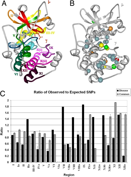
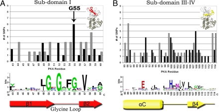
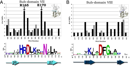
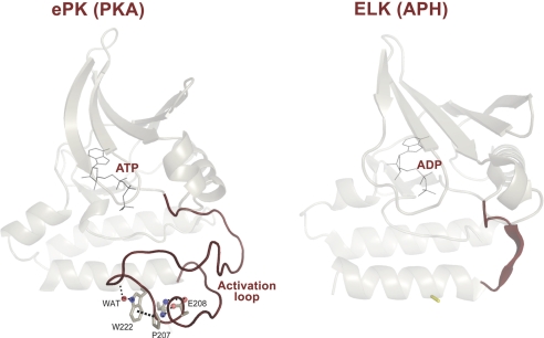
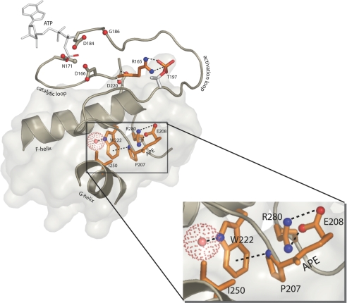
The catalytic domain of protein kinases harbors a large number of disease-causing single nucleotide polymorphisms (SNPs) and common or neutral SNPs that are not known or hypothesized to be associated with any disease. Distinguishing these two types of polymorphisms is critical in accurately predicting the causative role of SNPs in both candidate gene and genome-wide association studies. In this study, we have analyzed the structural location of common and disease-associated SNPs in the catalytic domain of protein kinases and find that, although common SNPs are randomly distributed within the catalytic core, known disease SNPs consistently map to regulatory and substrate binding regions. In particular, a buried side-chain network that anchors the flexible activation loop to the catalytic core is frequently mutated in disease patients. This network was recently shown to be absent in distantly related eukaryotic-like kinases, which lack an exaggerated activation loop and, presumably, are not regulated by phosphorylation.
Conflict of interest statement
The authors declare no conflict of interest.
Figures
References
-
- Knighton DR, et al. Crystal structure of the catalytic subunit of cyclic adenosine monophosphate-dependent kinase. Science. 1991;253:407–414. - PubMed
-
- Manning G, Whyte DB, Martinez R, Hunter T, Sudarsanam S. The protein kinase complement of the human genome. Science. 2002;298:1912–1934. - PubMed
-
- Ortutay C, Valiaho J, Stenberg K, Vihinen M. KinMutBase: A registry of disease-causing mutations in protein kinase domains. Hum Mutat. 2005;25:435–442. - PubMed
Publication types
MeSH terms
Substances
Grants and funding
- R01 HL070137-01/HL/NHLBI NIH HHS/United States
- U01 HL064777/HL/NHLBI NIH HHS/United States
- U19 AG023122/AG/NIA NIH HHS/United States
- HHMI/Howard Hughes Medical Institute/United States
- R01 GM019301/GM/NIGMS NIH HHS/United States
- R01 HL074730-02/HL/NHLBI NIH HHS/United States
- U01 HL064777-06/HL/NHLBI NIH HHS/United States
- U19 AG023122-01/AG/NIA NIH HHS/United States
- R01 HL074730/HL/NHLBI NIH HHS/United States
- R01 HL070137/HL/NHLBI NIH HHS/United States
- R37 GM019301/GM/NIGMS NIH HHS/United States
- GM19301/GM/NIGMS NIH HHS/United States
- 5 R01 HLMH065571-02/PHS HHS/United States
LinkOut - more resources
Full Text Sources
Medical
