. 2008 Jul 1;105(26):8914-9.
doi: 10.1073/pnas.0801559105. Epub 2008 Jun 25.

An epoxide hydrolase involved in the biosynthesis of an insect sex attractant and its use to localize the production site

Affiliations

An epoxide hydrolase involved in the biosynthesis of an insect sex attractant and its use to localize the production site

Mohatmed Abdel-Latief et al. Proc Natl Acad Sci U S A. .

Abstract

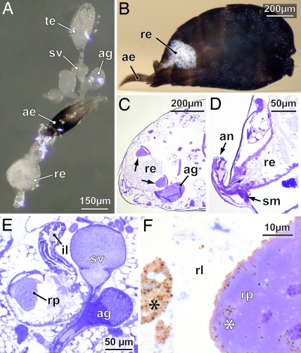
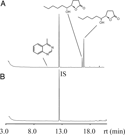
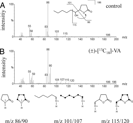
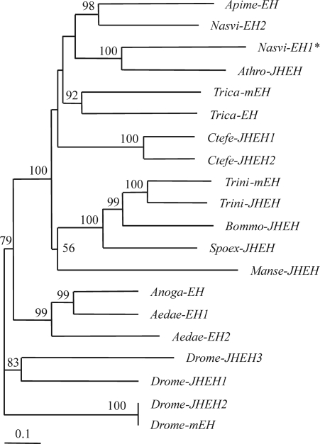
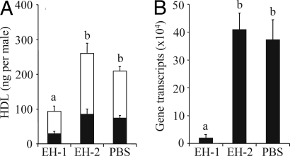
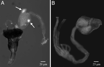
Epoxide hydrolases (EHs) are enzymes occurring in virtually any living organism. They catalyze the hydrolysis of epoxide containing lipids and are involved in crucial mechanisms, such as the detoxification of xenobiotics or the regulation of inflammation and blood pressure. Here, we describe a function of a putative EH gene in the biosynthesis of a sex attractant in the jewel wasp Nasonia vitripennis and use this gene to localize the site of pheromone production. Males of this parasitic wasp release a mixture of (4R,5R)-( threo-) and (4R,5S)-( erythro-)5-hydroxy-4-decanolide (HDL) to attract virgin females. Using a stable isotope labeled precursor, we demonstrated that vernolic acid ( erythro-12,13-epoxy-octadec-9Z-enoic acid) is converted by N. vitripennis males to threo-HDL. This suggested the involvement of an EH in hydrolyzing the fatty acid epoxide under inversion of the stereochemistry into the respective diol, which might be further processed by chain shortening and lactonization to HDL. We cloned a putative N. vitripennis EH gene (Nasvi-EH1) encoding 470 amino acids and localized its transcripts in the male rectal papillae by in situ RT-PCR. Chemical analyses and histological studies confirmed that males synthesize the sex attractant in the rectal vesicle and release it via the anal orifice. Involvement of Nasvi-EH1 in HDL biosynthesis was established by RNAi-mediated gene silencing. Injection of Nasvi-EH1 dsRNA into male abdomens inhibited pheromone biosynthesis by 55% and suppressed the targeted gene transcripts in the rectal vesicle by 95%.


Conflict of interest statement

The authors declare no conflict of interest.

Figures

Fig. 1.
Fig. 1.
Male reproductive system and hindgut of N. vitripennis. (A) Whole-mount of the dissected components used for localizing the pheromone gland. (B) View into a sagittally sliced abdomen in resin, showing the rectum filled with a whitish tubular accumulation (meconium). (C and D) Sagittal sections of the abdomen at two different levels, showing two rectal papillae (arrows) protruding into the rectal vesicle (C) and the opening of the rectum to the exterior via the anus, which can be dilated by rectal suspensory muscles (D). (E) Transverse section through rectal vesicle and right gonad, showing the rectal papilla flexibly attached to a strengthened muscular cap. (F) Detail of rectal papilla, showing vesicles (asterisks) both within the cone cells of the rectal papilla and aggregated in the lumen of the rectal vesicle. ae, aedeagus; ag, accessory gland; an, anus; il, ileum; re, rectum; rl, rectal lumen; rp, rectal papilla; sm, suspensory muscle (dilator); sv, seminal vesicle; te, testis.
Fig. 2.
Fig. 2.
Localization of the pheromone gland in the male abdomen. Total ion current chromatograms of dichloromethane extracts from rectal vesicle (A) and interior genitalia (testis, seminal vesicle, and accessory gland) (B) of a single N. vitripennis male. IS indicates the internal standard (20 ng of methyl undecanoate).
Fig. 3.
Fig. 3.
Mass spectral data for threo-HDL from N. vitripennis obtained after application of 0.1 μl of acetone (control) (A) and (±)-[13C18]-vernolic acid (B). Pairs of diagnostic ions indicating the incorporation of the labeled precursor are shown below. 13C-Atoms are represented by black dots.
Fig. 4.
Fig. 4.
Phylogenetic tree analysis of insect epoxide hydrolase genes. The asterisk indicates the putative EH from N. vitripennis (Nasvi-EH1). N, vitripennis (Nasvi-EH2), Apis mellifera (Apime-EH), A. rosae (Athro-JHEH), Manduca sexta (Manse-JHEH), Bombyx mori (Bommo-JHEH), Spodoptera exigua (Spoex-JHEH), Trichoplusia ni (Trini-mEH, and EH), Drosophila melanogaster (Drome-JHEH1, 2, and 3 and mEH), Aedes aegypti (Aedae-JHEH1, and 2), Ctenocephalides felis (Canfe-EH1, and 2), and Tribolium castaneum (Trica-EH, and mEH). The amino acid alignment (shown in Fig. S5) was carried out by using CLUSTAL W. The corrected distance tree was rooted by declaring the D. melanogaster (Drome-mEH) and its homolog (Drome-JHEH) as outgroup based on its role as homolog to insect JHEH (26). The neighbor-joining tree was produced by using the PHYLIP package and based on the consensus of 1,000 bootstrap replicates. Numbers above branches are the whole percentage values of 1,000 neighbor-joining bootstraps. Tree drawing was performed with the help of TreeView 1.6.6.
Fig. 5.
Fig. 5.
Effects of RNAi treatment on HDL titers of N. vitripennis males (threo-HDL, black proportion of columns; erythro-HDL, white proportion of columns) (A) and the quantity of targeted Nasvi-EH1 gene transcripts in male rectal vesicles (B) as estimated by quantitative real time RT-PCR analysis. For RNAi treatment, males were injected with dsRNA corresponding to Nasvi-EH1 (EH1), and control males were injected with dsRNA corresponding to Nasvi-EH2 (EH2) or 1× DEPC-PBS buffer (PBS). Lowercase letters indicate significant differences between treatments (one-way ANOVA, least significant difference test).
Fig. 6.
Fig. 6.
Tissue-specific localization of the N. vitripennis epoxide hydrolase gene (Nasvi-EH1), using whole-mount in situ RT-PCR analysis. (A) Nasvi-EH1 was localized in the male rectal papillae. (B) No expression signal was detected in females. Sites of localization are signed with arrows. The Nasvi-EH1 mRNAs were labeled by using Dig 11-dUTP and visualized by fluorescence in situ RT-PCR analysis, using HNPP/Fast Red TR.

Similar articles

Cited by

References

    1. Hardie J, Minks AK. Pheromones of Non-Lepidopteran Insects Associated with Agricultural Plants. Wallingford, CT: CABI; 1999.
    1. Wyatt TD. Pheromones and Animal Behaviour. Cambridge: Cambridge Univ Press; 2003.
    1. Ma PWK, Ramaswamy SB. Biology and ultrastructure of sex pheromone-producing tissue. In: Blomquist G, Vogt R, editors. Insect Pheromone Biochemistry and Molecular Biology. London: Elsevier; 2003. pp. 19–51.
    1. Tillman JA, Seybold SJ, Jurenka RA, Blomquist GJ. Insect pheromones—an overview of biosynthesis and endocrine regulation. Insect Biochem Mol Biol. 1999;29:481–514. - PubMed
    1. Jurenka RA. Insect pheromone biosynthesis. Top Curr Chem. 2004;239:97–132. - PubMed

Publication types

MeSH terms

Associated data

LinkOut - more resources