. 2008 Sep 1;112(5):1767-75.
doi: 10.1182/blood-2008-01-132068. Epub 2008 Jun 25.

Rac GTPase isoforms Rac1 and Rac2 play a redundant and crucial role in T-cell development

Affiliations

Rac GTPase isoforms Rac1 and Rac2 play a redundant and crucial role in T-cell development

Fukun Guo et al. Blood. .

Abstract

Rac GTPases have been implicated in the regulation of diverse functions in various blood cell lineages, but their role in T-cell development is not well understood. We have carried out conditional gene targeting to achieve hematopoietic stem cell (HSC)- or T-cell lineage-specific deletion of Rac1 or Rac1/Rac2 by crossbreeding the Mx-Cre or Lck-Cre transgenic mice with Rac1(loxp/loxp) or Rac1(loxp/loxp);Rac2(-/-) mice. We found that (1) HSC deletion of both Rac1 and Rac2 inhibited production of common lymphoid progenitors (CLPs) in bone marrow and suppressed T-cell development in thymus and peripheral organs, whereas deletion of Rac1 moderately affected CLP production and T-cell development. (2) T cell-specific deletion of Rac1 did not affect T-cell development, whereas deletion of both Rac1 and Rac2 reduced immature CD4(+)CD8(+) and mature CD4(+) populations in thymus as well as CD4(+) and CD8(+) populations in spleen. (3) The developmental defects of Rac1/Rac2 knockout T cells were associated with proliferation, survival, adhesion, and migration defects. (4) Rac1/Rac2 deletion suppressed T-cell receptor-mediated proliferation, IL-2 production, and Akt activation in thymocytes. Thus, Rac1 and Rac2 have unique roles in CLP production and share a redundant but essential role in later stages of T-cell development by regulating survival and proliferation signals.


Figures

Figure 1
Figure 1
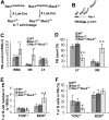
Gene targeting of Rac1 or Rac1/Rac2 in HSCs and the effect on peripheral blood and bone marrow cells. (A) Rac1 gene–targeted allele. The conditional Rac1 allele was generated by sandwiching exon 1 of Rac1 gene with 2 loxP sites. (B) Generation of Rac1 or Rac1/Rac2 knockout HSCs in mice. To produce Rac1- or Rac1/Rac2-deficient hematopoietic cells in mice, Rac1loxp/loxp or Rac1loxp/loxp/Rac2−/− mice were crossbred with Mx-Cre transgenic mice. Four to 5 doses of poly I:C injection of the mice resulted in efficient deletion of Rac1 gene in HSCs. (C) Expression of Rac1 in bone marrow, thymus, and spleen of Rac1 or Rac1/Rac2 Mx-Cre–targeted mice. Bone marrow cells, thymocytes, and splenocytes from WT-, Rac1-, or Rac1/Rac2-targeted mice were probed for Rac1 protein by anti-Rac1 blotting. Levels of β-actin were used as loading controls. (D) Deletion of Rac1/Rac2 but not Rac1 alone led to increased white blood cells (WBCs) and neutrophils (NEs) without apparent effect on lymphocyte numbers (LY) in peripheral blood. PB of various genotypes was analyzed by blood counting on a hematology analyzer. (E) PB of various genotypes was stained by biotinylated anti-CD3 or -B220 antibody followed by Streptavidin-Percp and analyzed by flow cytometry. (F) Deletion of Rac1 or Rac1/Rac2 reduced CLP numbers in bone marrow. Bone marrow cells were counted and stained for lineage markers with biotinylated antibodies against B220, CD3, CD4, CD8, Gr1, CD11b, and TER119. Subsequently, cells were stained with Streptavidin-Percp, anti–IL7R-APC-Cy7, anti–c-kit-APC, and anti–Sca1-PE. CLP was defined by the LinIL-7Rα+Sca1medc-kitmed-high phenotype. The number of CLP was calculated based on the total number of bone marrow cells and the percentage of CLP. n = 5 in each panel. *P < .05; **P < .01. Error bars represent SD.
Figure 2
Figure 2
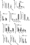
Deletion of Rac1 or Rac1/Rac2 in HSCs affects T-cell development and survival in thymus. (A) Thymic cellularity of Mx-Cre–targeted mice was determined in the indicated genotypes. Single-cell suspensions were prepared from freshly isolated thymus for quantification. (B) The numbers of T-cell subpopulations in thymocytes were determined by anti-CD4, -CD8, -CD44, and -CD25 staining and FACS analysis. (C) The numbers of DN thymocyte subpopulations were determined using additional anti-CD44 and anti-CD25 antibodies and subsequent FACS analysis. (D) The apoptotic cells in thymic T-cell subpopulations were determined by anti-CD4 and -CD8 and annexin V staining followed by flow cytometry. The results are representative of 2 experiments. n = 5 for each genotype. *P < .05; **P < .01. Error bars represent SD.
Figure 3
Figure 3
Deletion of Rac1 or Rac1/Rac2 in HSCs results in decreased T-lymphocyte cellularity in spleen that is associated with increased apoptosis. (A) Number of T cells in splenocytes was determined by staining the splenocytes with anti-CD4, -CD8, and -TCRβ antibodies followed by flow cytometry. (B) The apoptotic T-cell subpopulations in splenocytes were measured by the antibody and annexin V costaining followed by flow cytometry. The results are representative of 2 experiments. n = 5 for each genotype. **P < .01. Error bars represent SD.
Figure 4
Figure 4
Gene targeting of Rac1 or Rac1/Rac2 in the T-cell lineage and the effect on peripheral blood cells. (A) Generation of T cell–specific Rac1- or Rac1/Rac2-deficient mice. To produce Rac1- or Rac1/Rac2-deficient T lymphocytes, Rac1loxp/loxp or Rac1loxp/loxp/Rac2−/− mice were crossbred with Lck-Cre transgenic mice. (B) Expression of Rac1 in Lck-Cre–targeted thymocytes. WT, Rac1−/−, or Rac1−/−Rac2−/− thymocytes were probed for Rac1 protein expression by anti-Rac1 Western blotting. Levels of β-actin in each sample were probed as loading controls. (C) Deletion of Rac1/Rac2, but not Rac1 alone, led to increased WBC and NE counts but normal LY counts in peripheral blood. (D) Deletion of Rac1/Rac2 but not Rac1 by Lck-Cre targeting caused decreased LY percentage and increased NE percentage in peripheral blood cells. (E,F) The effects of Rac1 or Rac1/Rac2 deletion on T- and B-cell productions in peripheral blood. PB cells were stained for TCRβ or B220 and analyzed by flow cytometry. The T- and B-cell numbers and percentages in PB were presented in panels E and F, respectively. n = 7 for each genotype. *P < .05; **P < .01. Error bars represent SD.
Figure 5
Figure 5
Deletion of Rac1/Rac2, but not Rac1 alone, in T cells affects the development and proliferation/survival/adhesion/migration of thymocyte T cells. Lck-Cre–targeted mice were injected intraperitoneally with BrdU (100 μg/g body weight) 12 hours prior to harvesting thymus. Isolated thymocytes were quantified for the absolute number (A) and stained with anti-CD4, -CD8, -CD44, -CD25, -CD69, -HSA, -CD62L, and/or -BrdU antibodies or annexin V. The stained cells were subjected to flow cytometry analysis of the CD4/CD8 subpopulations (B), the expression level of HSA/CD62L in CD4+ or CD8+ SP thymocytes (C), the CD44/CD25 expression in CD4CD8 DN subpopulations (D), the CD69 level in CD4+CD8+ DP thymocytes (E), the proliferating cells (F), and the apoptotic cells (G) in various T-cell subpopulations. The adhesion to fibronectin and migration to SDF-1α of total thymocytes (H) or migration of CD4+ or CD8+ SP thymocyte to MIP-3β (I) was also determined. WT, n = 10; Rac1−/−, n = 6; and Rac1−/−Rac2−/−, n = 6. *P < .05; **P < .01. Error bars represent SD.
Figure 6
Figure 6
Deletion of Rac1/Rac2, but not Rac1 alone, in T cells results in decreased T-lymphocyte cellularity in spleen that is associated with decreased T-cell proliferation, adhesion, and migration and increased apoptosis. Lck-Cre–targeted mice were injected intraperitoneally with BrdU (100 μg/g body weight) 12 hours prior to spleen harvest. The isolated splenocytes were counted and stained with anti-CD4, -CD8, -TCRβ, and -BrdU antibodies or annexin V followed by flow cytometry analysis of the CD4/CD8 subpopulations (A), the proliferating cells (B), and the apoptotic cells (C) in various T-cell subpopulations. The purified splenic T cells were also assayed for adhesion to fibronectin and for migration toward SDF-1α (D). WT, n = 10; Rac1−/−, n = 6; and Rac1−/−Rac2−/−, n = 6. *P < .05; **P < .01. Error bars represent SD.
Figure 7
Figure 7
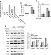
T cell–specific deletion of Rac1/Rac2 inhibits TCR-induced cell growth and signaling. Single-cell suspensions of thymocytes or splenic T cells were prepared from mice of the indicated genotypes. The cells were plated on 96-well plates at 2 × 10⁵/well in 100 μL culture with or without anti-TCR antibody or IL-2 (A-C). The medium was collected at day 2 from the thymocyte culture for IL-2 assay by ELISA (B), and the cell numbers were counted after a 3-day culture (A,C). Data are expressed as the fold of growth relative to the number of WT cells without stimulation. Error bars represent the standard deviations of 5 WT and 5 Rac1−/−Rac2−/− mice. *P < .05; **P < .01. (D) Western blotting was performed to assess the phosphorylation status of Akt, Erk, p38, and ZAP70 after a 3-minute TCR cross-linking of freshly isolated thymocytes. The quantitative results were shown as ratios between normalized phospho-Akt, -Erk, -p38, or -ZAP70 and total Akt, Erk, p38, or ZAP70, and are presented as means plus or minus SD from 5 WT and 5 Rac1−/−Rac2−/− mice. Rac1 expression was probed by anti-Rac1 blotting in parallel. Error bars represent SD.

Similar articles

Cited by

References

    1. Van Aelst L, D'Souza-Schorey C. Rho GTPases and signaling networks. Genes Dev. 1997;11:2295–2322. - PubMed
    1. Zheng Y. Dbl family guanine nucleotide exchange factors. Trends Biochem Sci. 2001;26:724–732. - PubMed
    1. Etienne-Manneville S, Hall A. Rho GTPases in cell biology. Nature. 2002;420:629–635. - PubMed
    1. Wang L, Zheng Y. Cell type specific function of Rho GTPases revealed by gene targeting in mice. Trends Cell Biol. 2007;17:58–64. - PubMed
    1. Hall A. Rho GTPases and the actin cytoskeleton. Science. 1998;279:509–514. - PubMed

Publication types

MeSH terms