Comparative Study
. 2008 May;15(5):437-47.
doi: 10.1177/1933719107314065.

Programming of human monocytes by the uteroplacental environment

Affiliations
Comparative Study

Programming of human monocytes by the uteroplacental environment

Ramsey H McIntire et al. Reprod Sci. 2008 May.

Abstract

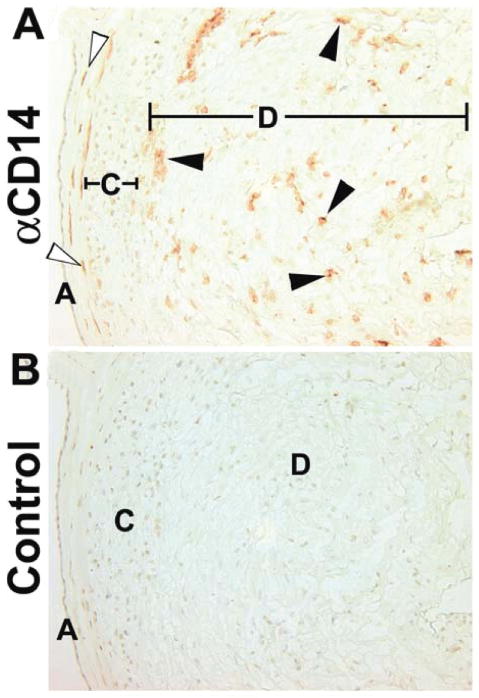
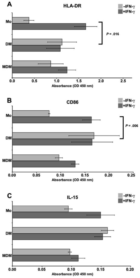
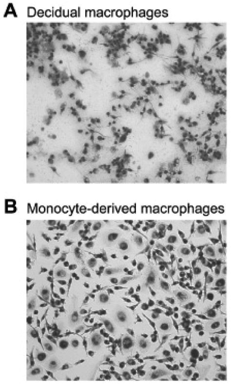
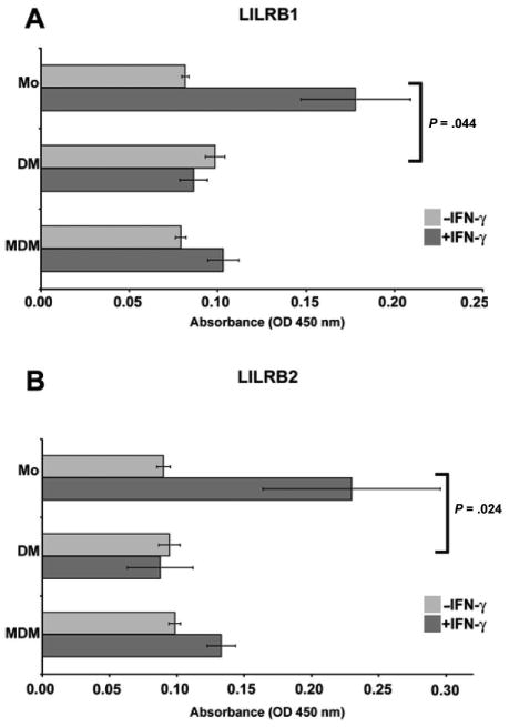
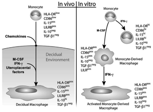
During human pregnancy, monocytes recruited to the uterus (decidua) are modified to promote immune defense and semiallogeneic pregnancy. The purpose of this study was to identify decidual factors involved in programming of monocytes into decidual macrophages by comparing the surface and secretory phenotypes of resting and interferon- gamma (IFN-gamma)-activated monocytes, unfractionated decidual cells, purified term decidual macrophages, and monocyte-derived macrophages. Surface markers for antigen presentation (HLA-DR, CD86), a membrane-bound cytokine interleukin (IL)-15, leukocyte immunoglobulin-like receptors (LILRB1, LILRB2), and secreted anti-inflammatory cytokines (transforming growth factor [TGF]-beta1 and IL-10) were assessed. The results demonstrate that differentiated, activated monocytes closely resemble but are not identical to decidual macrophages. In addition to differential IFN-gamma responsiveness, decidual macrophages were smaller than monocyte-derived macrophages and produced IL-10, which monocyte-derived macrophages did not. Only the unfractionated decidual cells secreted TGF-beta1. These results suggest that activation, differentiation, and decidual signals cooperate to program monocytes into the decidual macrophage phenotype.


Figures

Figure 1
Figure 1
Immunohistochemical localization of macrophages in term extraplacental membranes. (A) Anti-CD14. (B) Isotype matched control. Open arrows mark fetal macrophages between the amnion and chorion membranes. Closed arrows mark decidual macrophages close to the chorion membrane and scattered randomly through the decidua. A, amnion epithelium; C, chorion cytotrophoblast cell layer; D, decidua. Original magnifications, 200×.
Figure 2
Figure 2
Analysis of activation markers on monocytes, purified decidual macrophages, and monocyte-derived macrophages using cell enzyme-linked immunosorbent assay (ELISA). Antibodies used to obtain the results were (A) anti-HLA-DR, (B) anti-CD86, and (C) anti-IL-15. In each panel, markers on blood monocytes (Mo), purified decidual macrophages (DM), and monocyte-derived macrophages (MDM) cultured in the absence (−IFN-γ) or presence (+IFN-γ) of 100 U/mL interferon-γ (IFN-γ) for 48 hours are shown. OD indicates optical density. Data shown are the means of values obtained in at least 3 separate experiments ± SEM. Brackets and P values indicate statistical significance between fold changes in response to IFN-γ treatment.
Figure 3
Figure 3
Morphologic appearances of (A) decidual macrophages cultured for 48 hours in medium containing interferon-γ (IFN-γ) and (B) monocyte-derived macrophages, which had been cultured for 5 days in medium containing 150 IU/mL macrophage colony-stimulating factor followed by 48 hours in medium containing 100 U/mL IFN-γ. Original magnifications, 200×.
Figure 4
Figure 4
Analysis of HLA-G receptors on monocytes, purified decidual macrophages, and monocyte-derived macrophages by cell surface enzyme-linked immunosorbent assay. (A) Leukocyte immunoglobulin-like receptor (LILR)B1 and (B) LILRB2. Blood monocytes (Mo), purified decidual macrophages (DM), and monocyte-derived macrophages (MDM) were cultured in the absence (−IFN-γ) or presence (+IFN-γ) of 100 U/mL interferon-γ (IFN-γ) for 48 hours. OD indicates optical density. Data shown are the mean values obtained in 3 separate experiments ± SEM. Brackets and P values indicate statistical significance between fold changes in response to IFN-γ treatment.
Figure 5
Figure 5
Analysis of (A) HLA-DR, (B) CD86, and (C) IL-15 on resting (−IFN-γ) and activated (+IFN-γ) purified decidual macrophages and unfractionated decidual cells using a cell enzyme-linked immunosorbent assay. OD indicates optical density. Data shown are the mean values obtained in 3 separate experiments ± SEM. Brackets and P values indicate statistical significance between absorbance values regardless of interferon-γ (IFN-γ) treatment.
Figure 6
Figure 6
Schematic illustration showing marker expression of decidual macrophages (left panel) in comparison with in vitro–activated monocytes, differentiated monocytes, and monocytes that were both differentiated and activated (right panel). IFN, interferon; IL, interleukin; M-CSF, macrophage colony-stimulating factor; TGF, transforming growth factor.

Comment in

  • Macrophages and pregnancy.
    Mor G, Koga K. Mor G, et al. Reprod Sci. 2008 May;15(5):435-6. doi: 10.1177/1933719108317253. Reprod Sci. 2008. PMID: 18579852 No abstract available.

References

    1. Gordon S. Alternative activation of macrophages. Nat Rev Immunol. 2003;3:23–35. - PubMed
    1. Rutherford MS, Witsell A, Schook LB. Mechanisms generating functionally heterogeneous macrophages: chaos revisited. J Leukoc Biol. 1993;53:602–618. - PubMed
    1. Vince GS, Johnson PM. Immunobiology of human uteroplacental macrophages—friend and foe? Placenta. 1996;17:191–199. - PubMed
    1. Mor G, Abrahams VM. Potential role of macrophages as immunoregulators of pregnancy. Reprod Biol Endocrinol. 2003;1:119. - PMC - PubMed
    1. Hunt JS, Pollard JW. Macrophages in the uterus and placenta. Curr Top Microbiol Immunol. 1992;181:39–63. - PubMed

Publication types

MeSH terms