Molecular imaging-assisted optimization of hsp70 expression during laser-induced thermal preconditioning for wound repair enhancement
- PMID: 18580963
- PMCID: PMC3846389
- DOI: 10.1038/jid.2008.175
Molecular imaging-assisted optimization of hsp70 expression during laser-induced thermal preconditioning for wound repair enhancement
Abstract
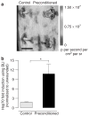
Patients at risk for impaired healing may benefit from prophylactic measures aimed at improving wound repair. Several photonic devices claim to enhance repair by thermal and photochemical mechanisms. We hypothesized that laser-induced thermal preconditioning would enhance surgical wound healing that was correlated with hsp70 expression. Using a pulsed diode laser (lambda=1.85 microm, tau(p)=2 ms, 50 Hz, H=7.64 mJ cm(-2)), the skin of transgenic mice that contain an hsp70 promoter-driven luciferase was preconditioned 12 hours before surgical incisions were made. Laser protocols were optimized in vitro and in vivo using temperature, blood flow, and hsp70-mediated bioluminescence measurements as benchmarks. Biomechanical properties and histological parameters of wound healing were evaluated for up to 14 days. Bioluminescent imaging studies indicated that an optimized laser protocol increased hsp70 expression by 10-fold. Under these conditions, laser-preconditioned incisions were two times stronger than control wounds. Our data suggest that this molecular imaging approach provides a quantitative method for optimization of tissue preconditioning and that mild laser-induced heat shock may be a useful therapeutic intervention prior to surgery.
Conflict of interest statement
The authors state no conflict of interest.
Figures


Similar articles
-
In vivo analysis of laser preconditioning in incisional wound healing of wild-type and HSP70 knockout mice with Raman spectroscopy.Lasers Surg Med. 2012 Mar;44(3):233-44. doi: 10.1002/lsm.22002. Epub 2012 Jan 24. Lasers Surg Med. 2012. PMID: 22275297 Free PMC article.
-
Assessing laser-tissue damage with bioluminescent imaging.J Biomed Opt. 2006 Jul-Aug;11(4):041114. doi: 10.1117/1.2339012. J Biomed Opt. 2006. PMID: 16965142
-
Expression of heat shock proteins 70 and 47 in tissues following short-pulse laser irradiation: assessment of thermal damage and healing.Med Eng Phys. 2013 Oct;35(10):1406-14. doi: 10.1016/j.medengphy.2013.03.011. Epub 2013 Apr 12. Med Eng Phys. 2013. PMID: 23587755
-
Assessment of cellular response to thermal laser injury through bioluminescence imaging of heat shock protein 70.Photochem Photobiol. 2004 Jan;79(1):76-85. Photochem Photobiol. 2004. PMID: 14974719
-
Can thermal lasers promote skin wound healing?Am J Clin Dermatol. 2003;4(1):1-12. doi: 10.2165/00128071-200304010-00001. Am J Clin Dermatol. 2003. PMID: 12477368 Review.
Cited by
-
In-vivo optical imaging of hsp70 expression to assess collateral tissue damage associated with infrared laser ablation of skin.J Biomed Opt. 2008 Sep-Oct;13(5):054066. doi: 10.1117/1.2992594. J Biomed Opt. 2008. PMID: 19021444 Free PMC article.
-
Temporal gene expression kinetics for human keratinocytes exposed to hyperthermic stress.Cells. 2013 Apr 10;2(2):224-43. doi: 10.3390/cells2020224. Cells. 2013. PMID: 24709698 Free PMC article.
-
THz irradiation inhibits cell division by affecting actin dynamics.PLoS One. 2021 Aug 2;16(8):e0248381. doi: 10.1371/journal.pone.0248381. eCollection 2021. PLoS One. 2021. PMID: 34339441 Free PMC article.
-
In vivo analysis of laser preconditioning in incisional wound healing of wild-type and HSP70 knockout mice with Raman spectroscopy.Lasers Surg Med. 2012 Mar;44(3):233-44. doi: 10.1002/lsm.22002. Epub 2012 Jan 24. Lasers Surg Med. 2012. PMID: 22275297 Free PMC article.
-
Postabdominoplasty Scar Improvement after a Single Session with an Automated 1210-nm Laser.Plast Reconstr Surg Glob Open. 2023 Mar 10;11(3):e4866. doi: 10.1097/GOX.0000000000004866. eCollection 2023 Mar. Plast Reconstr Surg Glob Open. 2023. PMID: 36910728 Free PMC article.
References
-
- Allain JC, Le Lous M, Cohen-Solal BS, Bazin S, Maroteaux P. Isometric tensions developed during the hydrothermal swelling of rat skin. Connect Tissue Res. 1980;7:127–33. - PubMed
-
- Baskaran H, Toner M, Yarmush ML, Berthiaume F. Poloxamer-188 improves capillary blood flow and tissue viability in a cutaneous burn wound. J Surg Res. 2001;101:56–61. - PubMed
-
- Beckham JT, Mackanos MA, Crooke C, Takahashi T, O’Connell-Rodwell C, Contag CH, et al. Assessment of cellular response to thermal laser injury through bioluminescence imaging of heat shock protein 70. Photochem Photobiol. 2004;79:76–85. - PubMed
-
- Beckham JT, Wilmink G. 2007 ASLMS abstracts—disclosure and FDA status. Lasers Surg Med. 2007;39:87–100. - PubMed
-
- Beere HM, Wolf BB, Cain K, Mosser DD, Mahboubi A, Kuwana T, et al. Heat-shock protein 70 inhibits apoptosis by preventing recruitment of procaspase-9 to the Apaf-1 apoptosome. Nat Cell Biol. 2000;2:469–75. - PubMed
Publication types
MeSH terms
Substances
Grants and funding
LinkOut - more resources
Full Text Sources
Other Literature Sources

