

Twenty-first century brain banking: practical prerequisites and lessons from the past: the experience of New York Brain Bank, Taub Institute, Columbia University
- PMID: 18581261
- PMCID: PMC2847415
- DOI: 10.1007/s10561-008-9079-y
Twenty-first century brain banking: practical prerequisites and lessons from the past: the experience of New York Brain Bank, Taub Institute, Columbia University
Abstract
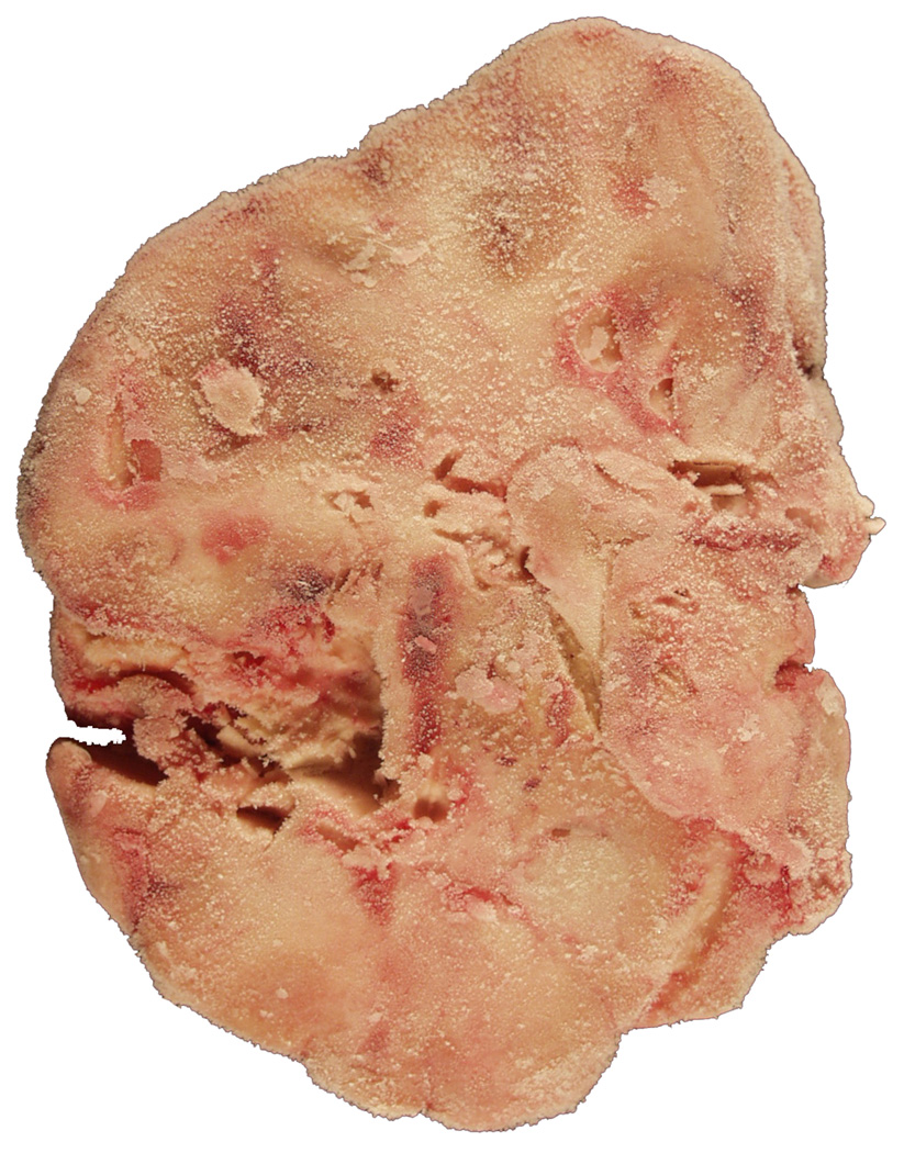
Generally accepted methods for processing postmortem brains are lacking, despite the efforts of pioneers in the field, and the growing awareness of the importance of brain banking for investigating the pathogenesis of illnesses unique to humans. Standardizing methods requires compromises, institutional or departmental mindset promoting collaboration, and the willingness to share ideas, information, and samples. A sound balance between competition and institutional interests is needed to best fulfill the tasks entrusted to health care institutions. Thus, a potentially widely accepted protocol design involves tradeoffs. We successfully integrated brain banking within the operation of the department of pathology. We reached a consensus whereby a brain can be utilized for diagnosis, research, and teaching. Thus, routing brains away from residency programs is avoided. The best diagnostic categorization possible is being secured and the yield of samples for research maximized. Thorough technical details pertaining to the actual processing of brains donated for research were recently published. Briefly, one-half of each brain is immersed in formalin for performing the neuropathologic evaluation, which is combined with the teaching task. The contralateral half is extensively dissected at the fresh state to obtain samples ready for immediate disbursement once categorized diagnostically. The samples are tracked electronically, which is crucial. This important tracking system is described separately in this issue. This report focuses on key lessons learned over the past 25 years of brain banking including successful solutions to originally unforeseen problems.
Figures


Similar articles
-
Twenty-first century brain banking. Processing brains for research: the Columbia University methods.Acta Neuropathol. 2008 May;115(5):509-32. doi: 10.1007/s00401-007-0311-9. Epub 2007 Nov 6. Acta Neuropathol. 2008. PMID: 17985145 Free PMC article.
-
The New York Brain Bank of Columbia University: practical highlights of 35 years of experience.Handb Clin Neurol. 2018;150:105-118. doi: 10.1016/B978-0-444-63639-3.00008-6. Handb Clin Neurol. 2018. PMID: 29496134 Review.
-
Electronic tracking of human brain samples for research.Cell Tissue Bank. 2008 Sep;9(3):217-27. doi: 10.1007/s10561-008-9078-z. Epub 2008 Jul 9. Cell Tissue Bank. 2008. PMID: 18612850 Free PMC article.
-
Twenty-first century brain banking: at the crossroads.Acta Neuropathol. 2008 May;115(5):493-6. doi: 10.1007/s00401-008-0363-5. Acta Neuropathol. 2008. PMID: 18347804
-
Brain banking in the United States.J Neuropathol Exp Neurol. 2003 Jul;62(7):715-22. doi: 10.1093/jnen/62.7.715. J Neuropathol Exp Neurol. 2003. PMID: 12901698 Review.
Cited by
-
Pathology and Biobanking.Turk Patoloji Derg. 2020;36(2):93-108. doi: 10.5146/tjpath.2020.01482. Turk Patoloji Derg. 2020. PMID: 32189322 Free PMC article. Review.
-
Elevated expression of the retrotransposon LINE-1 drives Alzheimer's disease-associated microglial dysfunction.Acta Neuropathol. 2024 Nov 27;148(1):75. doi: 10.1007/s00401-024-02835-6. Acta Neuropathol. 2024. PMID: 39604588 Free PMC article.
-
Epigenome signatures landscaped by histone H3K9me3 are associated with the synaptic dysfunction in Alzheimer's disease.Aging Cell. 2020 Jun;19(6):e13153. doi: 10.1111/acel.13153. Epub 2020 May 17. Aging Cell. 2020. PMID: 32419307 Free PMC article.
-
An International Survey of Brain Banking Operation and Characterization Practices.Biopreserv Biobank. 2016 Dec;14(6):464-469. doi: 10.1089/bio.2016.0003. Epub 2016 Jul 11. Biopreserv Biobank. 2016. PMID: 27399803 Free PMC article.
-
Lipocalin-2 Is a Key Regulator of Neuroinflammation in Secondary Traumatic and Ischemic Brain Injury.Neurotherapeutics. 2023 Apr;20(3):803-821. doi: 10.1007/s13311-022-01333-5. Epub 2022 Dec 12. Neurotherapeutics. 2023. PMID: 36508119 Free PMC article.
References
-
- Alafuzoff I, Parkkinen L, Al-Sarraf S, et al. Assessment of α-synuclein pathology: A study of the BrainNet Europe Consortium. Journal of Neuropathology and Experimental Neurology. 2008;67:125–143. - PubMed
-
- Alzheimer A. Über eigenartige Krankheitsfälle des späteren Alters. Zeitschrift für die gesamte Neurologie und Psychiatrie (Berlin) 1911;4:356–385.
-
- Aström K-E, Mancall EL, Richardson EP., Jr Progressive multifocal leuko-encephalopathy. A hitherto unrecognized complication of chronic lymphatic leukaemia and Hodgkin's disease. Brain. 1958;81:93–111. - PubMed
-
- Bertrand I, van Bogaert L. La sclérose latérale amyotrophique (anatomie pathologique) Revue Neurologique. 1925;1:779–806.
-
- Bird ED, Vonsattel J-P. The development of a brain bank. Journal of Neural Transmission [Suppl] 1993;39:17–23. - PubMed
Publication types
MeSH terms
Grants and funding
LinkOut - more resources
Full Text Sources
Miscellaneous
