. 2008 Jul 23;130(29):9251-3.
doi: 10.1021/ja801445p. Epub 2008 Jun 27.

Protein-protein interaction detection in vitro and in cells by proximity biotinylation

Affiliations

Protein-protein interaction detection in vitro and in cells by proximity biotinylation

Marta Fernández-Suárez et al. J Am Chem Soc. .

Abstract

We report a new method for detection of protein-protein interactions in vitro and in cells. One protein partner is fused to Escherichia coli biotin ligase (BirA), while the other protein partner is fused to BirA's "acceptor peptide" (AP) substrate. If the two proteins interact, BirA will catalyze site-specific biotinylation of AP, which can be detected by streptavidin staining. To minimize nonspecific signals, we engineered the AP sequence to reduce its intrinsic affinity for BirA. The rapamycin-controlled interaction between FKBP and FRB proteins could be detected in vitro and in cells with a signal to background ratio as high as 28. We also extended the method to imaging of the phosphorylation-dependent interaction between Cdc25C phosphatase and 14-3-3epsilon phosphoserine/threonine binding protein. Protein-protein interaction detection by proximity biotinylation has the advantages of low background, high sensitivity, small AP tag size, and good spatial resolution in cells.


Figures

Figure 1
Figure 1. Protein-protein interaction detection with biotin ligase and an engineered acceptor peptide substrate
(A) Detection scheme. Interaction between proteins A and B results in site-specific biotinylation of the acceptor peptide (AP) by biotin ligase (BirA). (B) Left: domain structures of constructs used for in vitro and cellular tests. Right: FKBP was fused to the original AP sequence as well as to 7 truncated variants of the AP with increased Km for BirA. The lysine biotinylation site is underlined. Placeholder amino acids inserted at truncation sites are colored green. (C) Comparison of AP truncation mutants in a live cell assay. HEK cells co-transfected with FRB-BirA and one of the FKBP fusions to a truncated AP (FKBP-AP′) were either treated with 100 nM rapamycin for 1 hour or left untreated. Biotin was then added for 1 minute before lysis and analysis by streptavidin western blot. The lower α-myc blot visualizes the myc tag in each FKBP-AP′ construct.
Figure 2
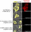
Figure 2. Imaging the FKBP-FRB interaction in cells
HEK cells were co-transfected with FRB-BirA and FKBP-AP(-3). After 3 hours incubation with 100 nM rapamycin, cells were labeled with biotin for 1 minute, then fixed and stained with streptavidin-Alexa Fluor 568 (red). Anti-HA staining (yellow) shows HA-FRB-BirA expression. DIC (differential interference contrast) images on the left show total cells in the field of view. Negative controls are shown with rapamycin omitted, an alanine mutation in AP(-3), and a K183R mutation in BirA that eliminates its activity.
Figure 3
Figure 3. Proximity biotinylation provides subcellular localization of protein-protein interactions in cells
TRex™-HEK cells were cotransfected with NLS-FRB-BirA and NLS-FKBP-AP(-3). NLS is a nuclear localization signal. Protein expression was induced with tetracycline for 24 hours. Cells were labeled with biotin as above, then fixed and stained with streptavidin-Alexa Fluor 647 (purple). Anti-HA staining (yellow) detects FRB expression, and anti-myc staining (red) detects total FKBP expression. Figure S5 shows results from imaging cells co-expressing untargeted FRB-BirA with NLS-FKBP-AP(-3), as well as cells co-expressing NLS-FRB-BirA with untargeted FKBP-AP(-3).
Figure 4
Figure 4. Imaging the interaction between Cdc25C and 14-3-3ε in cells
TRex™-HEK cells were co-transfected with mCherry-AP(-3)-Cdc25C and BirA-14-3-3ε, and protein expression was induced with tetracycline for 24 hours. Cells were then labeled with biotin for 1 minute, and fixed and stained with streptavidin-Alexa Fluor 647 (purple). Anti-HA staining (yellow) detects total HA-BirA-14-3-3ε expression, and mCherry fluorescence (red) highlights Cdc25C. Controls are shown with the noninteracting S216A Ccd25C mutant, and with BirA-14-3-3ε replaced by FRB-BirA.

References

    1. Truong K, Ikura M. Curr.Opin.Struct.Biol. 2001;11(5):573–578. - PubMed
    1. Remy I, Michnick SW. Methods Mol.Biol. 2004;261:411–426. - PubMed
    1. Michnick SW. Curr.Opin.Biotechnol. 2003;14(6):610–617. - PubMed
    1. Hu CD, Kerppola TK. Nat.Biotechnol. 2003;21(5):539–545. - PMC - PubMed
    1. Galarneau A, Primeau M, Trudeau LE, Michnick SW. Nat.Biotechnol. 2002;20(6):619–622. - PubMed

Publication types

MeSH terms