

The machinery for cell polarity, cell morphogenesis, and the cytoskeleton in the Basidiomycete fungus Ustilago maydis-a survey of the genome sequence
- PMID: 18582586
- PMCID: PMC2615052
- DOI: 10.1016/j.fgb.2008.05.012
The machinery for cell polarity, cell morphogenesis, and the cytoskeleton in the Basidiomycete fungus Ustilago maydis-a survey of the genome sequence
Abstract
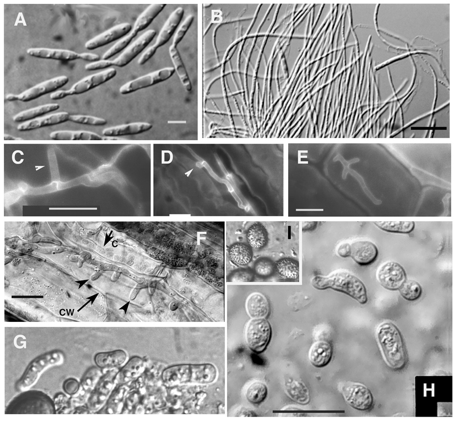
Ustilago maydis, a Basidiomycete fungus that infects maize, exhibits two basic morphologies, a yeast-like and a filamentous form. The yeast-like cell is elongated, divides by budding, and the bud grows by tip extension. The filamentous form divides at the apical cell and grows by tip extension. The repertoire of morphologies is increased during interaction with its host, suggesting that plant signals play an important role in generation of additional morphologies. We have used Saccharomyces cerevisiae and Schizosaccharomyces pombe genes known to play a role in cell polarity and morphogenesis, and in the cytoskeleton as probes to survey the U. maydis genome. We have found that most of the yeast machinery is conserved in U. maydis, albeit the degree of similarity varies from strong to weak. The U. maydis genome contains the machinery for recognition and interpretation of the budding yeast axial and bipolar landmarks; however, genes coding for some of the landmark proteins are absent. Genes coding for cell polarity establishment, exocytosis, actin and microtubule organization, microtubule plus-end associated proteins, kinesins, and myosins are also present. Genes not present in S. cerevisiae and S. pombe include a homolog of mammalian Rac, a hybrid myosin-chitin synthase, and several kinesins that exhibit more similarity to their mammalian counterparts. We also used the U. maydis genes identified in this analysis to search other fungal and other eukaryotic genomes to identify the closest homologs. In most cases, not surprisingly, the closest homolog is among filamentous fungi, not the yeasts, and in some cases it is among mammals.
Figures
References
-
- Akhmanova A, Hoogenraad CC. Microtubule plus-end-tracking proteins: mechanisms and functions. Curr. Opin. Cell Biol. 2005;17:47–54. - PubMed
-
- Banuett F. History of the mating types in Ustilago maydis. In: Heitman J, Kronstad J, Taylor J, Casselton LA, editors. Sex in Fungi: Molecular determination and evolutionary implications. Washington, DC: ASM press; 2007. pp. 351–375.
-
- Banuett F. Pathogenic development in Ustilago maydis: A progression of morphological transitions that results in tumor formation and teliospore production. In: Osiewacz HD, editor. Molecular Biology of Fungal Development. New York, Basel: Marcel Dekker; 2002. pp. 349–398.
-
- Banuett F. Genetics of Ustilago maydis, a fungal pathogen that induces tumors in maize. Annu. Rev. Genetics. 1995;29:179–208. - PubMed
Publication types
MeSH terms
Substances
Grants and funding
LinkOut - more resources
Full Text Sources
Molecular Biology Databases
Miscellaneous
