. 2008 Aug;173(2):385-99.
doi: 10.2353/ajpath.2008.070729. Epub 2008 Jun 26.

Transforming growth factor-beta stimulates intestinal epithelial focal adhesion kinase synthesis via Smad- and p38-dependent mechanisms

Affiliations

Transforming growth factor-beta stimulates intestinal epithelial focal adhesion kinase synthesis via Smad- and p38-dependent mechanisms

Mary F Walsh et al. Am J Pathol. 2008 Aug.

Abstract

Focal adhesion kinase (FAK) regulates cell migration, proliferation, and apoptosis. FAK protein is reduced at the edge of migrating gut epithelial sheets in vitro, but it has not been characterized in restitutive gut mucosa in vivo. Here we show that FAK and activated phospho-FAK (FAK(397)) immunoreactivity was lower in epithelial cells immediately adjacent to human gastric and colonic ulcers in vivo, but dramatically increased in epithelia near the ulcers, possibly reflecting stimulation by growth factors absent in vitro. Transforming growth factor (TGF)-beta, but not fibroblast growth factor, platelet-derived growth factor, or vascular endothelial growth factor, increased FAK levels in Caco-2 and IEC-6 cells. Epithelial immunoreactivity to TGF-beta and phospho-Smad3 was also higher near the ulcers, varying in parallel with FAK. The TGF-beta receptor antagonist SB431542 completely blocked TGF-beta-induced Smad2/3 and p38 activation in IEC-6 cells. SB431542, the p38 antagonist SB203580, and siRNA-mediated reduction of Smad2 and p38alpha prevented TGF-beta stimulation of both FAK transcription and translation (as measured via a FAK promoter-luciferase construct). FAK(397) levels were directly related to total FAK protein expression. Although gut epithelial motility is associated with direct inhibition of FAK protein adjacent to mucosal wounds, TGF-beta may increase FAK protein near but not bordering mucosal ulcers via Smad2/3 and p38 signals. Our results show that regulation of FAK expression may be as important as FAK phosphorylation in critically influencing gut epithelial cell migration after mucosal injury.


Figures
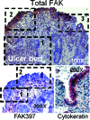
Figure 1
Figure 1
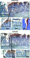
Benign human gastric ulcers exhibit three zones of FAK and phospho-FAK (FAK397) immunoreactivity in vivo. Top: In this representative ulcer, expression of total FAK (purple/brown stain) is extremely low immediately adjacent to the ulcer bed (zone 1), greatly elevated at the healing edge (zone 2), and again much lower in the distant mucosa (zone 3). Bottom left: Similarly, immunostaining for phosphorylated FAK397, lower at the tip of the migrating epithelial sheet (zone 1), is increased in the healing mucosa (zone 2). Staining is most prominent at the basal aspect of the cells where they contact the basement membrane matrix and where FAK would be expected to be functional. Bottom right: Intensity of staining in the cytokeratin control was similar throughout. Original magnifications: ×100 (top); ×200 (bottom).
Figure 2
Figure 2
Colonic ulcers show a similar pattern of FAK immunoreactivity. A: Intensity of staining for total FAK shows an increasing gradient from low near the ulcer bed (zone 1) to high in the region adjacent to it (zone 2) (top). This transition (arrow) is demonstrated more clearly when the area is magnified (bottom left). Also magnified, zone 3, away from the ulcer bed, exhibits very low staining for total FAK (middle) while staining for the control cytokeratin remains even throughout (bottom right). B: FAK397 immunostaining is similarly reduced at the ulcer margin (zone 1) with a transition to higher staining in the adjacent zone (zone 2). Again, the transition zone is clearly seen under magnification (zone 2, bottom left), and FAK397 staining is reduced in cells distal to the ulcer (bottom right). In both the total and FAK397 micrographs, zone 3, although not contiguous to zones 1 and 2 was taken from the same section but removed slightly because mucosal sloughing obscured the transition between zones 2 and 3. Arrows point to enlarged transition areas in both the total and FAK397 immunostains. Hematoxylin was used as a counterstain in both the gastric and colonic ulcer micrographs, representative of 41 human ulcers. Original magnifications: ×100 (A, B; top), ×200 (A, B; bottom).
Figure 3
Figure 3
A: TGF-β increases immunoreactive FAK protein levels in vitro. FAK protein abundance, expressed as the ratio of total FAK to α-tubulin and as a percentage of untreated control in cells treated with TGF-β (1 ng/ml), bFGF (5 ng/ml), PDGF-BB (2.5 ng/ml), or VEGF (5 ng/ml) for 24 hours, is significantly higher only in TGF-β-treated IEC-6 (left bars) and Caco-2 (right bars) cells (*P < 0.05, n = 3 to 5). B: Total and activated FAK are enhanced to a similar degree after 24 hours of exposure to TGF-β. Densitometric analyses of total FAK and FAK397:α-tubulin ratios as a percentage of their respective control are depicted for IEC-6 (left bars) and Caco-2 (right bars) cells (*P < 0.02, n = 4 for each). Representative blots are shown above the bars.
Figure 4
Figure 4
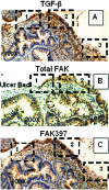
In vivo, the pattern of colonic ulcer epithelial cell TGF-β immunoreactivity mirrors that of FAK. A: In another colonic ulcer, TGF-β immunoreactivity exhibits a similar distribution: low intensity in cells at the migrating front (zone 1), higher intensity in zone 2, and lower intensity distal to the ulcer (zone 3). For comparison, total FAK immunostaining in an immediately adjacent section reveals the same low-high-lower pattern of staining intensity in epithelial cells bordering the ulcer (B), and FAK397 immunoreactivity is similarly low in the ulcer zone and elevated beyond it (C). Original magnifications: ×100 (A, C); ×200 (B).
Figure 5
Figure 5
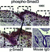
The pattern of colonic epithelial cell phospho-Smad3 (pSmad3) immunoreactivity mirrors that of FAK and TGF-β. Top: Immunoreactivity for pSmad3 is low immediately adjacent to the ulcer bed (zone 1), elevated at the healing edge (zone 2), and intermediate in intensity in the distant mucosa (zone 3). Bottom left: Higher magnification of the transition area from zone 1 at the tip of the migrating epithelial sheet to the healing mucosa (zone 2). Bottom right: A mouse isotype control showing lack of color development in epithelial cells. Original magnifications: ×100 (top, bottom right); ×200 (bottom left).
Figure 6
Figure 6
In vitro, TGF-β enhances FAK immunoreactivity in motile IEC-6 cells. Left: IEC-6 cells migrating across a linear wound were immunostained for total FAK (tFAK, top) or TGF-β (bottom). Lines delineate the location of the scrape; direction of migration is from left to right. Nuclei were counterstained with hematoxylin. tFAK and TGF-β immunoreactivity is decreased in motile cells: cytoplasmic immunostaining intensity is lower in cells migrating across the scrape than in those behind the front for either tFAK (top left) or TGF-β (bottom left). Right: TGF-β treatment increases tFAK immunoreactivity in motile cells, as evidenced by the level of cytoplasmic staining in the expanding monolayer (top). Intensity of staining for ERK as control (bottom) is essentially unchanged in motile cells (representative of three separate experiments). Original magnifications, ×10.
Figure 7
Figure 7
TGF-β enhances FAK protein expression at the level of transcription in Caco-2 and IEC-6 cells. A: Activity of the FAK promoter-luciferase construct, normalized to Renilla and expressed as percentage of the respective nontreated control, is significantly higher in IEC-6 and Caco-2 cells 24 hours after TGF-β treatment (*P = 0.007 versus nontreated respective control, n = 4 for each). B: FAK mRNA levels are higher in IEC-6 cells as early as 2 hours after treatment with TGF-β. RT-PCR results, normalized to the 18S control, are expressed as fold increase over nontreated control cells (*P < 0.05, n = 5).
Figure 8
Figure 8
TGF-β-mediated Smad2/3 activation is blocked by a TGF-β receptor antagonist but not by p38MAPK inhibition. IEC-6 cells were pretreated with SB431542 (0.5 μmol/L) or SB203580 (10 μmol/L) for 1 hour before TGF-β exposure, also for 1 hour. The TGF-β-induced rise in Smad2/3 serine phosphorylation (∼15-fold; *P = 0.0004 versus DMSO vehicle-treated control) is totally abolished by SB431542. The TGF-β receptor blocker also significantly lowers basal Smad2/3 phosphorylation (**P = 0.0001). The p38 inhibitor, SB203580, has no effect on Smad2/3 activation (***P = 0.0001 versus SB203580 control). Bars represent densitometric analysis of five separate experiments; a representative Western blot is shown above. In this and subsequent figures, results are presented as percentage of control but statistics were calculated using the original densitometric ratios of the protein of interest and its respective GAPDH or α-tubulin control.
Figure 9
Figure 9
TGF-β receptor antagonism or p38MAPK inhibition blocks TGF-β-stimulated p38 activation. A: p38 phosphorylation, significantly increased by TGF-β (*P = 0.05 versus DMSO control, n = 4), is abolished by pretreatment with SB431542. SB203580, known to block p38 activity at a step beyond phosphorylation has no effect (**P = 0.001 versus SB203580). HSP27 accumulation was therefore used as a surrogate index of p38 activation/inhibition. B: TGF-β-stimulated accumulation of HSP27 (*P = 0.006 versus DMSO control, n = 5) is completely abrogated by either SB431542 or SB203580. Densitometric analysis is shown in the bars; representative Western blots are shown above.
Figure 10
Figure 10
Smad2/3 or p38 signal inhibition abolishes TGF-β-mediated FAK protein expression. A: A 24-hour exposure to TGF-β increases immunoreactive FAK protein levels in IEC-6 cells (135 ± 9% compared to DMSO-treated controls, *P = 0.001). Pretreatment with SB431542, a TGF-β receptor blocker, or SB203580 to block p38 activation, negates this effect. B: FAK397 levels are correspondingly elevated by TGF-β exposure and similarly blocked by either SB431542 or SB203580. Bars represent densitometric analysis of 6 to 12 individual experiments; typical Western blots for total FAK or FAK397, and α-tubulin, are shown above. C: IEC-6 cells transfected with the FAK-promoter-luciferase construct exhibit a fourfold greater luciferase activity over the empty pGL2 vector and a further increase (*P = 0.008) on exposure to TGF-β. Neither SB431542 nor SB203580 has an effect on basal FAK promoter activity; both inhibitors block the response to TGF-β. Bars represent luciferase activity, normalized to that of the Renilla control and expressed as fold increase over the pGL2 empty vector (n = 4).
Figure 11
Figure 11
Lowering Smad2 and p38α protein expression with specific siRNA also abolishes TGF-β-mediated FAK protein expression. A: Introduction of Smad2 siRNA into IEC-6 cells decreased Smad2 protein expression to 23 ± 5% of that in the nontargeting siRNA (NT1) treated control (P = 0.0003, not shown). This reduction in Smad2 protein completely inhibits the TGF-β-induced increase in FAK expression. B: p38α siRNA decreased p38α protein (to 15 ± 3%) and total p38 protein (to 27 ± 5%) levels compared to the NT1 siRNA-treated controls (P = 0.008 versus NT1, not shown). Lowering p38 expression also blocks the TGF-β effect. Densitometric analysis of total FAK, expressed as a ratio of α-tubulin and as a percentage of the NT1 control is shown in the bars. Representative Western blots for FAK, Smad2, and p38α and their respective α-tubulin or GAPDH controls are shown above the corresponding bars.
Figure 12
Figure 12
TGF-β-stimulated p38 phosphorylation is independent of Smad2/3 activation. p38 phosphorylation is enhanced equally in cells treated with nontargeting siRNA (NT1, P = 0.002) and specific siRNA directed at Smad2 (siSmad2, P = 0.01) when compared to their respective non-TGF-β-treated controls.
Figure 13
Figure 13
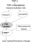
Schematic diagram illustrating TGF-β-induced FAK protein induction by parallel Smad2/3 and p38 signal pathways.

Similar articles

Cited by

References

    1. Burridge K, Nuckolls G, Otey C, Pavalko F, Simon K, Turner C. Actin-membrane interaction in focal adhesions. Cell Differ Dev. 1990;32:337–342. - PubMed
    1. Schaller MD, Borgman CA, Cobb BS, Vines RR, Reynolds AB, Parsons JT. pp125FAK a structurally distinctive protein-tyrosine kinase associated with focal adhesions. Proc Natl Acad Sci USA. 1992;89:5192–5196. - PMC - PubMed
    1. Gilmore AP, Romer LH. Inhibition of focal adhesion kinase (FAK) signaling in focal adhesions decreases cell motility and proliferation. Mol Biol Cell. 1996;7:1209–1224. - PMC - PubMed
    1. Salazar EP, Rozengurt E. Bombesin and platelet-derived growth factor induce association of endogenous focal adhesion kinase with Src in intact Swiss 3T3 cells. J Biol Chem. 1999;274:28371–28378. - PubMed
    1. Yu CF, Basson MD. Matrix-specific FAK and MAPK reorganization during Caco-2 cell motility. Microsc Res Tech. 2000;51:191–203. - PubMed

Publication types

MeSH terms