Review
. 2008 Aug;173(2):301-10.
doi: 10.2353/ajpath.2008.070752. Epub 2008 Jun 26.

Nucleolus, ribosomes, and cancer

Affiliations
Review

Nucleolus, ribosomes, and cancer

Lorenzo Montanaro et al. Am J Pathol. 2008 Aug.

Abstract

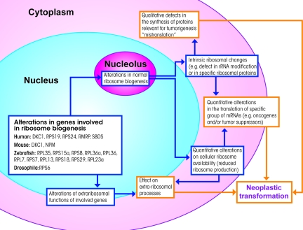
The complex aspects linking the nucleolus and ribosome biogenesis to cancer are reviewed here. The available evidence indicates that the morphological and functional changes in the nucleolus, widely observed in cancer tissues, are a consequence of both the increased demand for ribosome biogenesis, which characterizes proliferating cells, and the changes in the mechanisms controlling cell proliferation. In fact, the loss or functional changes in the two major tumor suppressor proteins pRB and p53 cause an up-regulation of ribosome biogenesis in cancer tissues. In this context, the association in human carcinomas of nucleolar hypertrophy with bad prognoses is worthy of note. Further, an increasing amount of data coming from studies on both hepatitis virus-induced chronic liver diseases and a subset of rare inherited disorders, including X-linked dyskeratosis congenita, suggests an active role of the nucleolus in tumorigenesis. Both an up-regulation of ribosome production and changes in the ribosome structure might causally contribute to neoplastic transformation, by affecting the balance of protein translation, thus altering the synthesis of proteins that play an important role in the genesis of cancer.


Figures

Figure 1
Figure 1
a: Human breast cancer cells cultured in vitro visualized using the contrast phase microscope. The nucleoli appear as highly refractive bodies. b: Histological section of human colon carcinoma stained with H&E. The nucleoli are intensely stained by eosin. c: Electron microscope visualization of the nucleolus of a TG cell (established cell line from a human fallopian tube cancer) after uranium and lead staining. Three fibrillar centers are present (asterisks), with the closely associated dense fibrillar component (f). The granular component (g) is organized in cord-like structures. d: TG cell, after specific AgNOR protein staining, at the electron microscopy level. Only the fibrillar centers and the associated dense fibrillar component are stained. e: Same preparation as in d, but visualized with light microscopy. The silver-stained structures appeared as well-defined, darkly stained dots clustered in the nucleolus. f and g: Histological sections of two human breast carcinomas, stained for the AgNOR proteins. The nucleoli are specifically stained. Note the small size on the nucleoli in the case reported in f, which was characterized by a good prognosis, and the larger size of nucleoli in the case reported in g, characterized by a fatal outcome. Scale bars: 12 μm (a); 18 μm (b); 0.3 μm (c); 1.25 μm (d); 3.25 μm (e); 20 μm (f, g).
Figure 2
Figure 2
Simplified diagram of the control mechanisms of cell proliferation and ribosome biogenesis by pRB and p53. Inactive pRB is linked to UBF and E2Fs. When the cell enters the cell cycle, phosphorylation of pRB by cyclin-dependent kinases 4 and 2 occurs, freeing UBF and the E2Fs. UBF, together with SL1, binds to the rDNA promoter, thus allowing rDNA transcription by RNA polymerase I (Pol I). The E2Fs bind to their target genes, thus allowing DNA duplication and cell cycle progression. DNA damage stabilizes p53, which blocks ribosome biogenesis both by directly binding to SL1 and by hindering pRB phosphorylation through the inhibitory action of p21 on Cdk4 and 2. Cdk4 may be also constrained by p16Ink4a. Ribosome biogenesis may also be inhibited by Arf, which binds to SL1 and stabilizes p53.
Figure 3
Figure 3
Schematic representation of the different cellular events through which altered ribosome biogenesis might contribute to neoplastic transformation. Alterations in the genes involved in ribosome biogenesis may generate intrinsic altered ribosomes (eg, hypopseudouridylated ribosomes in X-linked dyskeratosis congenita) characterized by an altered capacity for translating mRNAs coding for proteins associated with tumorigenesis. Alternatively, altered ribosome biogenesis may be associated with quantitative changes in cellular ribosome availability, which could influence translation of the genes involved in neoplastic transformation as well. It should also be considered that most of the proteins involved in ribosome biogenesis have extra-ribosomal functions that may support tumorigenesis independently of ribosome function and mRNA translation.

Similar articles

Cited by

References

    1. Pianese G. Beitrag zur Histologie und Aetiologie der Carcinoma. Histologische und experimentelle Untersuchungen. Beitr Pathol Anat Allgem Pathol. 1896;142:1–193.
    1. Busch H, Smetana K. New York: Academic Press,; The Nucleolus. 1970
    1. Hadjiolov AA. The nucleolus and ribosome biogenesis. Vienna: Springer Verlag,; 1985:pp 1–267.
    1. Derenzini M, Ploton D. Interphase nucleolar organizer regions in cancer cells. Int Rev Exp Pathol. 1991;32:149–192. - PubMed
    1. Conlon I, Raff M. Size control in animal development. Cell. 1999;96:235–244. - PubMed

Publication types

MeSH terms