. 2008 Aug 29;283(35):23895-902.
doi: 10.1074/jbc.M804069200. Epub 2008 Jun 26.

The basis for selective E1-E2 interactions in the ISG15 conjugation system

Affiliations

The basis for selective E1-E2 interactions in the ISG15 conjugation system

Larissa A Durfee et al. J Biol Chem. .

Abstract

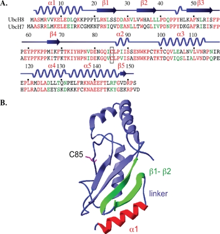
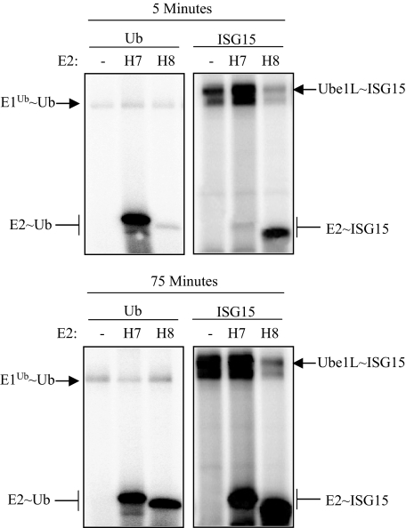
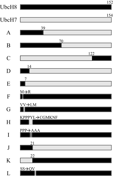
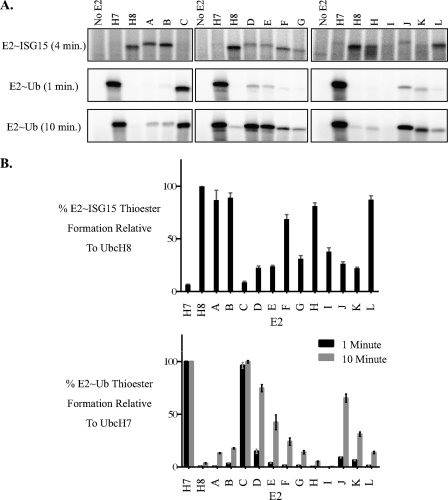
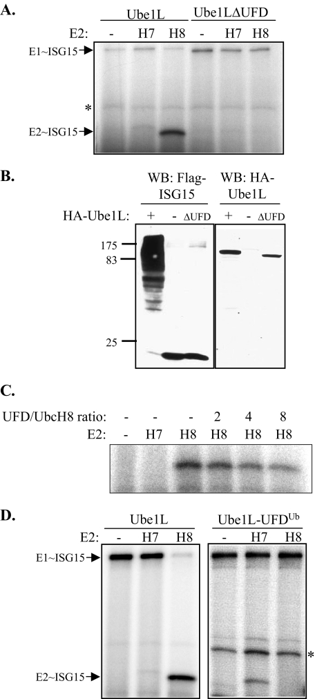
E1 and E2 enzymes coordinate the first steps in conjugation of ubiquitin (Ub) and ubiquitin-like proteins (Ubls). ISG15 is an interferon-alpha/beta-induced Ubl, and the E1 and E2 enzymes for ISG15 conjugation are Ube1L and UbcH8, respectively. UbcH7 is the most closely related E2 to UbcH8, yet it does not function in ISG15 conjugation in vivo, while both UbcH7 and UbcH8 have been reported to function in Ub conjugation. Kinetic analyses of wild-type and chimeric E2s were performed to determine the basis for preferential activation of UbcH8 by Ube1L and to determine whether UbcH8 is activated equally well by Ube1L and E1(Ub) (Ube1). K(m) determinations confirmed the strong preference of Ube1L for UbcH8 over UbcH7 (a 29-fold K(m) difference), similar to the preference of E1(Ub) for UbcH7 over UbcH8 (a 36-fold K(m) difference). Thioester assays of chimeric E2s identified two structural elements within residues 1-39 of UbcH8 that play a major role in defining Ube1L-UbcH8 specificity: the alpha1-helix and the beta1-beta2 region. The C-terminal ubiquitin fold domain (UFD) of Ube1L was required for transfer of ISG15 to UbcH8 and for binding of Ube1L to UbcH8. Replacement of the Ube1L UFD with that from E1(Ub) resulted in preferential transfer of ISG15 to UbcH7. Together, these results indicate that Ube1L discriminates between UbcH8 and closely related Ub E2s based on specific interactions between the Ube1L UFD and determinants within the N-terminal region of UbcH8.


Figures

FIGURE 1.
FIGURE 1.
A, alignment of the UbcH8 and UbcH7 sequences, with secondary structure elements of UbcH8 indicated. Numbering is according to UbcH8 residues. The active site cysteine residues are boxed. Residues in red represent identical residues, green represent similar residues. B, structure of UbcH8 (PDB 1WZW, Footnote 4). The α1-helix (red) and β1-β2 region (green) are indicated, along with the linker connecting these elements and the active site cysteine (C85, pink).
FIGURE 2.
FIGURE 2.
E1-E2 thioester assays with wild-type UbcH7 and UbcH8. Thioester complex formation was analyzed after incubation with E1Ub or Ube1L for either 5 min (top panel) or 75 min (bottom panel) with wild-type UbcH7 or UbcH8. The Ub and ISG15 were labeled with 32P and thioester adducts were detected by autoradiography.
FIGURE 3.
FIGURE 3.
Schematic of chimeric and mutant E2 proteins. UbcH8 sequences are shown in black and UbcH7 sequences are shown in gray. Numbering at chimera junctions represents the first residue (if the chimera contains UbcH8 in its C terminus) or the last residue (if the chimera contains UbcH8 in its N terminus) of the UbcH8 sequence present in the chimera. Specific amino acid changes are shown for some chimeras and mutants.
FIGURE 4.
FIGURE 4.
In vitro Ub and ISG15 thioester assays with chimeric E2s. A, equivalent amounts of the indicated E2 proteins were incubated with [32P]ISG15 and Ube1L for 4 min (top panel), [32P]Ub and E1Ub (DEAE-purified) for 1 min (middle panel), or [32P]Ub and E1Ub (DEAE-purified) for 10 min (bottom panel). Reaction products were analyzed by SDS-PAGE without reducing agent. B, E2 thioester adducts were quantitated and are represented as a percentage relative to UbcH8 (for ISG15 thioesters; upper panel) or relative to UbcH7 (for Ub thioesters; lower panel).
FIGURE 5.
FIGURE 5.
The UFD of Ube1L is required for the interaction with UbcH8. A, [32P]ISG15 was incubated for 10 min with Ube1L or Ube1LΔUFD, with the indicated E2 proteins, and reactions were analyzed by SDS-PAGE without reducing agent. An E2-independent background band is indicated (*). B, ISG15 conjugation in transfected 293 cells. 293 cells were transfected with plasmids expressing 3× FLAG-ISG15, UbcH8, Herc5, and either HA-Ube1L, no Ube1L, or HA-Ube1LΔUFD. Cell extracts were prepared and analyzed by immunoblotting with anti-FLAG antibody to detect ISG15 conjugates (left panel). Expression of HA-Ube1L and HA-Ube1LΔUFD was confirmed using anti-HA antibody (right panel). C, purified UFD of Ube1L is a competitive inhibitor of UbcH8∼ISG15 thioester formation. UbcH8 thioester formation was analyzed as in A, with increasing amounts of purified UFD protein present in the reaction (expressed as the molar ratio of UFD to UbcH8 protein). D, a chimeric Ube1L protein containing the UFD of E1Ub (Ube1L-UFDUb) preferentially transfers ISG15 to UbcH7. [32P]ISG15 was incubated with Ube1L or Ube1L-UFDUb and either no E2, UbcH7, or UbcH8. Reaction products were analyzed by SDS-PAGE without reducing agent. An E2-independent background band is indicated (*).

Similar articles

Cited by

References

    1. Pickart, C. M. (2001) Annu. Rev. Biochem. 70503 -533 - PubMed
    1. Melchior, F. (2000) Annu. Rev. Cell Dev. Biol. 16591 -626 - PubMed
    1. Scheffner, M., Nuber, U., and Huibregtse, J. M. (1995) Nature 37381 -83 - PubMed
    1. Kerscher, O., Felberbaum, R., and Hochstrasser, M. (2006) Annu. Rev. Cell Dev. Biol. 22 159-180 - PubMed
    1. Zhao, C., Beaudenon, S. L., Kelley, M. L., Waddell, M. B., Yuan, W., Schulman, B. A., Huibregtse, J. M., and Krug, R. M. (2004) Proc. Natl. Acad. Sci. U. S. A. 1017578 -7582 - PMC - PubMed

Publication types

MeSH terms