

doi: 10.1093/nar/gkn386.
Epub 2008 Jun 26.
A protein-DNA docking benchmark
Affiliations
- PMID: 18583363
- PMCID: PMC2504314
- DOI: 10.1093/nar/gkn386
Item in Clipboard
A protein-DNA docking benchmark
Nucleic Acids Res.
2008 Aug.
Abstract
We present a protein-DNA docking benchmark containing 47 unbound-unbound test cases of which 13 are classified as easy, 22 as intermediate and 12 as difficult cases. The latter shows considerable structural rearrangement upon complex formation. DNA-specific modifications such as flipped out bases and base modifications are included. The benchmark covers all major groups of DNA-binding proteins according to the classification of Luscombe et al., except for the zipper-type group. The variety in test cases make this non-redundant benchmark a useful tool for comparison and development of protein-DNA docking methods. The benchmark is freely available as download from the internet.
Figures
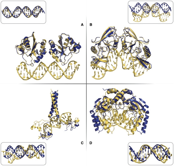
Illustration of ‘easy’ (interface RMSD < 2.0 Å), ‘intermediate’ (2.0 Å ≤ interface RMSD < 5.0 Å) and ‘difficult’ (interface RMSD ≥ 5.0 Å) test cases from the protein–DNA benchmark. ‘Easy’ test case: the Papillomavirus replication initiation domain E-1 (PDB id 1ksy) (interface RMSD = 1.6 Å) (A). ‘Intermediate’ test case: the intron-encoded homing endonuclease I-PPOI complex (PDB id 1a73) (interface RMSD = 4.3 Å) (B). ‘Difficult’ test cases: the proline utilization transcription activator (PDB id 1zme) (interface RMSD = 5.8 Å) (C) and the PVUII endonuclease complex (PDB id 1eyu) (interface RMSD = 6.8 Å) (D). The bound form of the complex is shown in yellow and the unbound protein in blue. The bound- and canonical B-form DNA structures are shown as insets to highlight the conformational changes in the DNA.
References
-
- van Dijk AD, Boelens R, Bonvin AM. Data-driven docking for the study of biomolecular complexes. FEBS J. 2005;272:293–312. - PubMed
-
- Janin J. The targets of CAPRI rounds 6-12. Proteins. 2007;69:699–703. - PubMed
-
- Rhodes D, Schwabe JW, Chapman L, Fairall L. Towards an understanding of protein-DNA recognition. Phil. Trans. Roy. Soc. Lond. 1996;351:501–509. - PubMed
-
- Mintseris J, Wiehe K, Pierce B, Anderson R, Chen R, Janin J, Weng Z. Protein-Protein Docking Benchmark 2.0: an update. Proteins. 2005;60:214–216. - PubMed
