. 2008 Sep;105(3):907-14.
doi: 10.1152/japplphysiol.00103.2008. Epub 2008 Jun 26.

Exercise training initiated after the onset of diabetes preserves myocardial function: effects on expression of beta-adrenoceptors

Affiliations

Exercise training initiated after the onset of diabetes preserves myocardial function: effects on expression of beta-adrenoceptors

Keshore R Bidasee et al. J Appl Physiol (1985). 2008 Sep.

Abstract

The present study was undertaken to assess cardiac function and characterize beta-adrenoceptor subtypes in hearts of diabetic rats that underwent exercise training (ExT) after the onset of diabetes. Type 1 diabetes was induced in male Sprague-Dawley rats using streptozotocin. Four weeks after induction, rats were randomly divided into two groups. One group was exercised trained for 3 wk while the other group remained sedentary. At the end of the protocol, cardiac parameters were assessed using M-mode echocardiography. A Millar catheter was also used to assess left ventricular hemodynamics with and without isoproterenol stimulation. beta-Adrenoceptors were assessed using Western blots and [(3)H]dihydroalprenolol binding. After 7 wk of diabetes, heart rate decreased by 21%, fractional shortening by 20%, ejection fraction by 9%, and basal and isoproterenol-induced dP/dt by 35%. beta(1)- and beta(2)-adrenoceptor proteins were reduced by 60% and 40%, respectively, while beta(3)-adrenoceptor protein increased by 125%. Ventricular homogenates from diabetic rats bound 52% less [(3)H]dihydroalprenolol, consistent with reductions in beta(1)- and beta(2)-adrenoceptors. Three weeks of ExT initiated 4 wk after the onset of diabetes minimized cardiac function loss. ExT also blunted loss of beta(1)-adrenoceptor expression. Interestingly, ExT did not prevent diabetes-induced reduction in beta(2)-adrenoceptor or the increase of beta(3)-adrenoceptor expression. ExT also increased [(3)H]dihydroalprenolol binding, consistent with increased beta(1)-adrenoceptor expression. These findings demonstrate for the first time that ExT initiated after the onset of diabetes blunts primarily beta(1)-adrenoceptor expression loss, providing mechanistic insights for exercise-induced improvements in cardiac function.


Figures

Fig. 1.
Fig. 1.
Representative echocardiograms (top) and cardiac parameters (table at bottom) of sedentary control, sedentary streptozotocin (STZ)-diabetic, exercise-trained (ExT) control, and ExT STZ-diabetic rats. For this, animals were lightly anesthetized with a cocktail containing ketamine (100 mg/ml) and acepromazine (10 mg/ml). Three loops of M-mode were captured for each animal, and data were averaged for 7–8 animals/group. Values shown are means ± SE. LVEDD, left ventricular end-diastolic diameter; LVESD, left ventricular end-systolic diameter; bpm, beats/min. *Significantly different from sedentary control (P < 0.05). **Significantly different from sedentary STZ-diabetic group (P < 0.05).
Fig. 2.
Fig. 2.
A: representative left ventricular (in vivo) pressure recordings of hearts from sedentary control, sedentary STZ-diabetic, ExT control, and ExT STZ-diabetic rats. Animals (7–8 animals/group) were lightly anesthetized with Inactin (20 mg/kg ip), and an F-2 micromanometer-tipped catheter (Millar Instruments, Houston, TX) was inserted via the right carotid artery into the left ventricle. B: mean values of left ventricular (LV) pressure. *Significantly different from sedentary control (P < 0.05). **Significantly different from sedentary STZ-diabetic (P < 0.05).
Fig. 3.
Fig. 3.
A: representative recordings of rates of left ventricular pressure changes (±dP/dt) in hearts from sedentary control, sedentary STZ-diabetic, ExT control, and ExT STZ-diabetic rats. Animals (7–8 animals per group) were lightly anesthetized with Inactin (20 mg/kg ip), and an F-2 micromanometer-tipped catheter (Millar Instruments) was inserted via the left carotid artery into the left ventricle. Data were obtained using Powerlab data-acquisition system (ADInstuments, Colorado Springs, CO). B: mean values for ±dP/dt. Iso, isoproterenol. *Significantly different from sedentary controls (P < 0.05). **Significantly different from sedentary STZ-diabetic (P < 0.05).
Fig. 4.
Fig. 4.
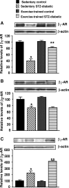
Representative Western blots showing steady state levels of β1- (A), β2- (B), and β3-adrenoceptors (AR) (C) and corresponding β-actin in left ventricular tissues from sedentary control, sedentary STZ-diabetic, ExT control, and ExT STZ-diabetic rat hearts. For this, 30 or 50 μg of membranes was used. Values n graphs represent the average data obtained from >5 separate preparations from each group analyzed in duplicate. *Significantly different from sedentary control (P < 0.05). **Significantly different from sedentary diabetic (P < 0.05).
Fig. 5.
Fig. 5.
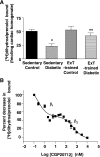
A: shows the ability of membranes prepared from sedentary control, sedentary STZ-diabetic, ExT control, and ExT STZ-diabetic rat hearts to bind [3H]dihydroalprenolol. Values in graphs represent the average data from >5 separate preparations from each group analyzed in duplicate. *Significantly different from sedentary control (P < 0.05). **Significantly different from sedentary diabetic (P < 0.05). B: displacement [3H]dihydroalprenolol binding assays were used to determine relative levels of β1- and β2-adrenoceptors in rat ventricular tissues using the β1-adrenoceptor selective agonist CGP-20712. Graph represents the average of >5 separate experiments done in duplicate (membranes were prepared from sedentary control ventricular tissues).

References

    1. Ades PA, Green NM, Coello CE. Effects of exercise and cardiac rehabilitation on cardiovascular outcomes. Cardiol Clin 21: 435–448, 2003. - PubMed
    1. Altan VM, Arioglu E, Guner S, Ozcelikay AT. The influence of diabetes on cardiac beta-adrenoceptor subtypes. Heart Fail Rev 12: 58–65, 2007. - PubMed
    1. American Diabetes Association. Complications of Diabetes in the United States.(http://www.diabetes.org/diabetes-statistics/complications.jsp).
    1. Arch JR β3-Adrenoceptor agonists: potential, pitfalls and progress. Eur J Pharmacol 440: 99–107, 2002. - PubMed
    1. Baldwin KM Effects of chronic exercise on biochemical and functional properties of the heart. Med Sci Sports Exerc 17: 522–528, 1985. - PubMed

Publication types

MeSH terms