

Assessment of dural arteriovenous fistulas of the cavernous sinuses on 3D dynamic MR angiography
- PMID: 18583402
- PMCID: PMC8118780
- DOI: 10.3174/ajnr.A1187
Assessment of dural arteriovenous fistulas of the cavernous sinuses on 3D dynamic MR angiography
Abstract
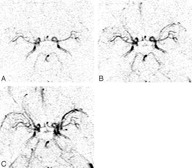
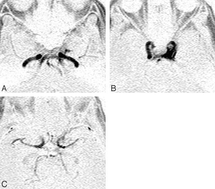
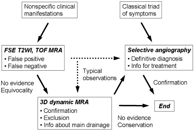
Background and purpose: Flow voids within the cavernous sinuses and/or certain venous drainage on spin-echo MR imaging and time-of-flight (TOF) flow enhancement on MR angiography (MRA) have indicated high-velocity shunt flow and have been used for screening patients with dural arteriovenous fistulas (DAVFs) of the cavernous sinuses. In this investigation, the capabilities of 3D dynamic MRA as a flow-independent approach and those of conventional MR imaging techniques were compared with selective angiography for the diagnosis of DAVFs of the cavernous sinuses.
Materials and methods: This retrospective study involved 18 patients with angiographically proved DAVFs of the cavernous sinuses and 12 control subjects. Sixteen partially overlapping sequential MR images were acquired on contrast-enhanced 3D dynamic MRA between the petrosal bone and the orbital roof. Two experienced observers blinded to the clinical data and results of angiography independently graded 3D dynamic MRA, fast spin-echo T2-weighted imaging (FSE T2WI), and TOF MRA.
Results: The average area under the receiver operating characteristic curve values and interobserver kappa scores for the diagnosis of DAVFs on 3D dynamic MRA, FSE T2WI, and TOF MRA were 0.99, 0.89, and 0.95; and 0.92, 0.71, and 0.73, respectively. Those for the diagnosis of anterior, posterior, and retrograde cortical venous drainage on 3D dynamic MRA were 0.72, 0.95, and 0.81; and 0.56, 0.50, and 0.49, respectively.
Conclusion: In this small series, screening 3D dynamic MRA directly demonstrates DAVFs of the cavernous sinuses and has improved diagnostic capability.
Figures
Similar articles
-
Evaluation of Intracranial Dural Arteriovenous Fistulas: Comparison of Unenhanced 3T 3D Time-of-flight MR Angiography with Digital Subtraction Angiography.Magn Reson Med Sci. 2015;14(4):285-93. doi: 10.2463/mrms.2014-0120. Epub 2015 May 19. Magn Reson Med Sci. 2015. PMID: 25994036
-
MR angiography of dural arteriovenous fistulas: diagnosis and follow-up after treatment using a time-resolved 3D contrast-enhanced technique.AJNR Am J Neuroradiol. 2007 May;28(5):877-84. AJNR Am J Neuroradiol. 2007. PMID: 17494662 Free PMC article. Clinical Trial.
-
The anterior medullary-anterior pontomesencephalic venous system and its bridging veins communicating to the dural sinuses: normal anatomy and drainage routes from dural arteriovenous fistulas.Neuroradiology. 2008 Dec;50(12):1013-23. doi: 10.1007/s00234-008-0433-3. Epub 2008 Jul 18. Neuroradiology. 2008. PMID: 18636248
-
MR angiography of the spine and spinal cord.Top Magn Reson Imaging. 2003 Dec;14(6):444-60. doi: 10.1097/00002142-200312000-00003. Top Magn Reson Imaging. 2003. PMID: 14872165 Review.
-
Spontaneous closure of non-cavernous sinus dural arteriovenous fistulas: A case series and systematic review of the literature.J Neuroradiol. 2022 Jan;49(1):94-100. doi: 10.1016/j.neurad.2020.09.002. Epub 2020 Sep 9. J Neuroradiol. 2022. PMID: 32918945
Cited by
-
Vessel-Selective 4D-MRA Using Superselective Pseudocontinuous Arterial Spin-Labeling with Keyhole and View-Sharing for Visualizing Intracranial Dural AVFs.AJNR Am J Neuroradiol. 2022 Mar;43(3):368-375. doi: 10.3174/ajnr.A7426. Epub 2022 Mar 3. AJNR Am J Neuroradiol. 2022. PMID: 35241425 Free PMC article.
-
Bilateral Low-Flow Type-D Dural Carotid-Cavernous Fistula: Diagnosis and Treatment with 3D Time-of-Flight Magnetic Resonance Angiography.Am J Case Rep. 2024 Mar 20;25:e942833. doi: 10.12659/AJCR.942833. Am J Case Rep. 2024. PMID: 38504435 Free PMC article.
-
Evaluation of dural arteriovenous fistulas of cavernous sinus before and after endovascular treatment using time-resolved MR angiography.Neurosurg Rev. 2010 Apr;33(2):217-22; discussion 222-3. doi: 10.1007/s10143-010-0246-9. Epub 2010 Feb 25. Neurosurg Rev. 2010. PMID: 20182900
-
Detection and grading of dAVF: prospects and limitations of 3T MRI.Eur Radiol. 2012 Feb;22(2):429-38. doi: 10.1007/s00330-011-2268-2. Epub 2011 Sep 21. Eur Radiol. 2012. PMID: 21932162
-
Evaluation of dural arteriovenous fistulas with 4D contrast-enhanced MR angiography at 3T.AJNR Am J Neuroradiol. 2010 Jan;31(1):80-5. doi: 10.3174/ajnr.A1898. Epub 2009 Oct 15. AJNR Am J Neuroradiol. 2010. PMID: 19833802 Free PMC article.
References
-
- Barrow DL, Spector RH, Braun IF, et al. Classification and treatment of spontaneous carotid-cavernous sinus fistulas. J Neurosurg 1985;62:248–56 - PubMed
-
- Halbach VV, Higashida RT, Hieshima GB, et al. Dural fistulas involving the cavernous sinus: results of treatment in 30 patients. Radiology 1987;163:437–42 - PubMed
-
- Chen YW, Jeng JS, Liu HM, et al. Carotid and transcranial color-coded duplex sonography in different types of carotid-cavernous fistula. Stroke 2000;31:701–06 - PubMed
-
- Stiebel-Kalish H, Setton A, Nimii Y, et al. Cavernous sinus dural arteriovenous malformations: patterns of venous drainage are related to clinical signs and symptoms. Ophthalmology 2002;109:1685–91 - PubMed
MeSH terms
LinkOut - more resources
Full Text Sources