

Differentiation between classic and atypical meningiomas with use of diffusion tensor imaging
- PMID: 18583409
- PMCID: PMC8118781
- DOI: 10.3174/ajnr.A1170
Differentiation between classic and atypical meningiomas with use of diffusion tensor imaging
Abstract
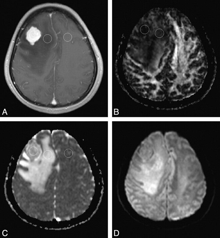
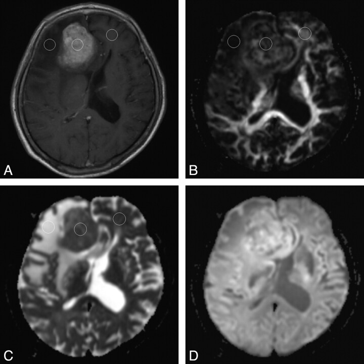
Background and purpose: The differentiation between classic and atypical meningiomas may have implications in preoperative planning but may not be possible on the basis of conventional MR imaging. Our hypothesis was that classic and atypical meningiomas have different patterns of intratumoral water diffusion that will allow for differentiation between them.
Materials and methods: Preoperative diffusion tensor imaging (DTI) was performed in 12 classic and 12 atypical meningiomas. Signal intensity of solid-enhancing tumor regions on diffusion-weighted trace images and apparent diffusion coefficient (ADC) and fractional anisotropy (FA) maps was assessed. Regions of interest (ROIs) were placed in solid-enhancing regions, peritumoral edema, and contralateral normal-appearing white matter (NAWM) to measure tensor metrics including major (lambda(1)), intermediate (lambda(2)) and minor eigenvalues (lambda(3)) and FA and ADC values. Distribution of tensor shapes within enhancing tumors was calculated for all tumors. Differences between classic and atypical meningiomas in tumor signal intensity, intratumoral and peritumoral tensor metrics, as well as tensor shapes distribution were statistically analyzed.
Results: A significantly greater proportion of atypical meningiomas were isointense and hypointense on ADC maps (P = .007). Classic meningiomas had significantly lower FA (P = .012), higher ADC (P = .011), greater lambda(2) (P = .020) and lambda(3) (P = .003). There was significantly more spherical diffusion in classic than in atypical meningiomas (P = .020). All diffusion tensor metrics for peritumoral edema of the 2 tumor groups did not differ.
Conclusion: DTI showed that intratumoral microscopic water motion is less organized in classic than in atypical meningiomas. This feature may allow for noninvasive differentiation between classic and atypical meningiomas.
Figures
References
-
- Bondy M, Ligon BL. Epidemiology and etiology of intracranial meningiomas: a review. J Neurooncol 1996;29:197–205 - PubMed
-
- Willis J, Smith C, Ironside JW, et al. The accuracy of meningioma grading: a 10-year retrospective audit. Neuropathol Appl Neurobiol 2005;31:141–49 - PubMed
-
- Modha A, Gutin PH. Diagnosis and treatment of atypical and anaplastic meningiomas: a review. Neurosurgery 2005;57:538–50 - PubMed
-
- Demaerel P, Wilms G, Lammens M, et al. Intracranial meningiomas: correlation between MR imaging and histology in fifty patients. J Comput Assist Tomogr 1991;15:45–51 - PubMed
-
- Perry A, Louis DN, Scheithauer BW, et al. Meningiomas. In: Louis DN, Ohgaki H, Wiestler OD, et al, eds. WHO Classification of Tumours of the Central Nervous System. Lyon: IARC Press;2007. :164–72
MeSH terms
LinkOut - more resources
Full Text Sources
Other Literature Sources
Medical