

Tumor suppression by p53 in the absence of Atm
- PMID: 18583527
- PMCID: PMC2680228
- DOI: 10.1158/1541-7786.MCR-07-2009
Tumor suppression by p53 in the absence of Atm
Abstract
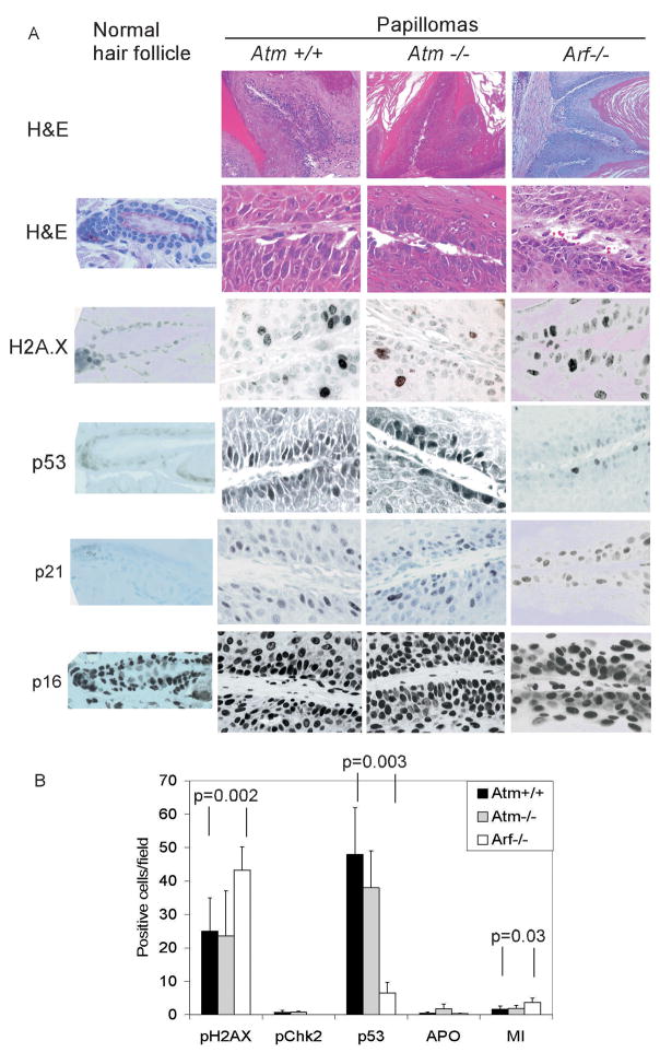
Oncogenes can induce p53 through a signaling pathway involving p19/Arf. It was recently proposed that oncogenes can also induce DNA damage, and this can induce p53 through the Atm DNA damage pathway. To assess the relative roles of Atm, Arf, and p53 in the suppression of Ras-driven tumors, we examined susceptibility to skin carcinogenesis in 7,12-dimethylbenz(a)anthracene/12-O-tetradecanoylphorbol-13-acetate (TPA)-treated Atm- and p53-deficient mice and compared these results to previous studies on Arf-deficient mice. Mice with epidermal-specific deletion of p53 showed increased papilloma number and progression to malignant invasive carcinomas compared with wild-type littermates. In contrast, Atm-deficient mice showed no increase in papilloma number, growth, or malignant progression. gamma-H2AX and p53 levels were increased in both Atm(+/+) and Atm(-/-) papillomas, whereas Arf(-/-) papillomas showed much lower p53 expression. Thus, although there is evidence of DNA damage, signaling through Arf seems to regulate p53 in these Ras-driven tumors. In spontaneous and radiation-induced lymphoma models, tumor latency was accelerated in Atm(-/-)p53(-/-) compound mutant mice compared with the single mutant Atm(-/-) or p53(-/-) mice, indicating cooperation between loss of Atm and loss of p53. Although p53-mediated apoptosis was impaired in irradiated Atm(-/-) lymphocytes, p53 loss was still selected for during lymphomagenesis in Atm(-/-) mice. In conclusion, in these models of oncogene- or DNA damage-induced tumors, p53 retains tumor suppressor activity in the absence of Atm.
Figures


Similar articles
-
Induction of p53 renders ATM-deficient mice refractory to hepatocarcinogenesis.Gastroenterology. 2010 Mar;138(3):1155-65.e1-2. doi: 10.1053/j.gastro.2009.11.008. Epub 2009 Nov 14. Gastroenterology. 2010. PMID: 19919837
-
Functional interaction of H2AX, NBS1, and p53 in ATM-dependent DNA damage responses and tumor suppression.Mol Cell Biol. 2005 Jan;25(2):661-70. doi: 10.1128/MCB.25.2.661-670.2005. Mol Cell Biol. 2005. PMID: 15632067 Free PMC article.
-
p19Arf suppresses growth, progression, and metastasis of Hras-driven carcinomas through p53-dependent and -independent pathways.PLoS Biol. 2004 Aug;2(8):E242. doi: 10.1371/journal.pbio.0020242. Epub 2004 Aug 17. PLoS Biol. 2004. PMID: 15314658 Free PMC article.
-
p53: guardian of the genome and policeman of the oncogenes.Cell Cycle. 2007 May 2;6(9):1006-10. doi: 10.4161/cc.6.9.4211. Epub 2007 May 28. Cell Cycle. 2007. PMID: 17457049 Review.
-
The ATM protein kinase and cellular redox signaling: beyond the DNA damage response.Trends Biochem Sci. 2012 Jan;37(1):15-22. doi: 10.1016/j.tibs.2011.10.002. Epub 2011 Nov 11. Trends Biochem Sci. 2012. PMID: 22079189 Free PMC article. Review.
Cited by
-
ATM and p53 in aging and cancer: a double-edged sword in genomic integrity.Biogerontology. 2025 May 5;26(3):102. doi: 10.1007/s10522-025-10249-4. Biogerontology. 2025. PMID: 40323544 Review.
-
Functional kinomics identifies candidate therapeutic targets in head and neck cancer.Clin Cancer Res. 2014 Aug 15;20(16):4274-88. doi: 10.1158/1078-0432.CCR-13-2858. Clin Cancer Res. 2014. PMID: 25125259 Free PMC article.
-
Doxorubicin enhances nucleosome turnover around promoters.Curr Biol. 2013 May 6;23(9):782-7. doi: 10.1016/j.cub.2013.03.043. Epub 2013 Apr 18. Curr Biol. 2013. PMID: 23602475 Free PMC article.
-
Synthetic lethal kinases in Ras/p53 mutant squamous cell carcinoma.Oncogene. 2022 Jun;41(24):3355-3369. doi: 10.1038/s41388-022-02330-w. Epub 2022 May 10. Oncogene. 2022. PMID: 35538224
-
RMP/URI inhibits both intrinsic and extrinsic apoptosis through different signaling pathways.Int J Biol Sci. 2019 Oct 15;15(12):2692-2706. doi: 10.7150/ijbs.36829. eCollection 2019. Int J Biol Sci. 2019. PMID: 31754340 Free PMC article.
References
-
- Banin S, Moyal L, Shieh S, Taya Y, Anderson CW, Chessa L, Smorodinsky NI, Prives C, Reiss Y, Shiloh Y, Ziv Y. Enhanced phosphorylation of p53 by ATM in response to DNA damage. Science. 1998;281:1674–1677. - PubMed
-
- Barlow C, Brown KD, Deng CX, Tagle DA, Wynshaw-Boris A. Atm selectively regulates distinct p53-dependent cell-cycle checkpoint and apoptotic pathways. Nature Genet. 1997;17:453–456. - PubMed
-
- Barlow C, Hirotsune S, Paylor R, Liyanage M, Eckhaus M, Collins F, Shiloh Y, Crawley JN, Ried T, Tagle D, Wynshaw-Boris A. Atm-deficient mice: a paradigm of ataxia telangiectasia. Cell. 1996;86:159–171. - PubMed
-
- Bartkova J, Horejsi Z, Koed K, Kramer A, Tort F, Zieger K, Guldberg P, Sehested M, Nesland JM, Lukas C, Orntoft T, Lukas J, Bartek J. DNA damage response as a candidate anti-cancer barrier in early human tumorigenesis. Nature. 2005;434:864–870. - PubMed
-
- Bartkova J, Rezaei N, Liontos M, Karakaidos P, Kletsas D, Issaeva N, Vassiliou LV, Kolettas E, Niforou K, Zoumpourlis VC, Takaoka M, Nakagawa H, Tort F, Fugger K, Johansson F, Sehested M, Andersen CL, Dyrskjot L, Orntoft T, Lukas J, Kittas C, Helleday T, Halazonetis TD, Bartek J, Gorgoulis VG. Oncogene-induced senescence is part of the tumorigenesis barrier imposed by DNA damage checkpoints. Nature. 2006;444:633–637. - PubMed
Publication types
MeSH terms
Substances
Grants and funding
LinkOut - more resources
Full Text Sources
Molecular Biology Databases
Research Materials
Miscellaneous
