. 2008 Aug;147(4):1735-49.
doi: 10.1104/pp.108.122226. Epub 2008 Jun 26.

The PRA1 gene family in Arabidopsis

Affiliations

The PRA1 gene family in Arabidopsis

Claire Lessa Alvim Kamei et al. Plant Physiol. 2008 Aug.

Abstract

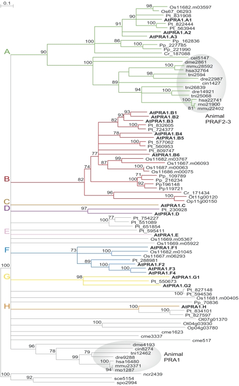
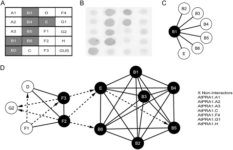
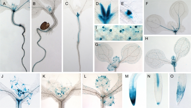
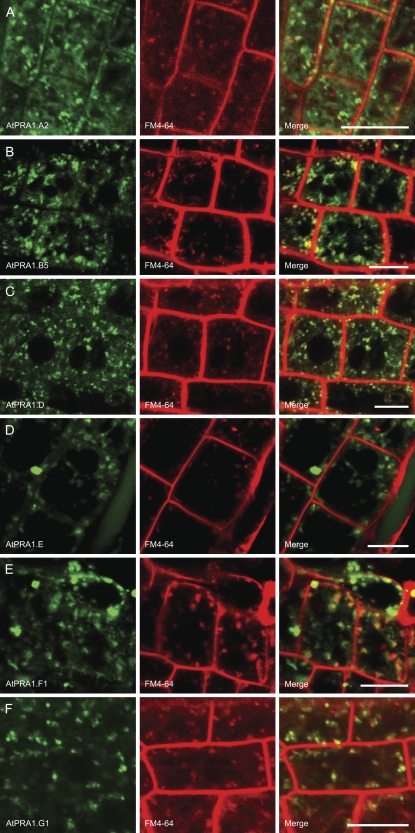
Prenylated Rab acceptor 1 (PRA1) domain proteins are small transmembrane proteins that regulate vesicle trafficking as receptors of Rab GTPases and the vacuolar soluble N-ethylmaleimide-sensitive factor attachment receptor protein VAMP2. However, little is known about PRA1 family members in plants. Sequence analysis revealed that higher plants, compared with animals and primitive plants, possess an expanded family of PRA1 domain-containing proteins. The Arabidopsis (Arabidopsis thaliana) PRA1 (AtPRA1) proteins were found to homodimerize and heterodimerize in a manner corresponding to their phylogenetic distribution. Different AtPRA1 family members displayed distinct expression patterns, with a preference for vascular cells and expanding or developing tissues. AtPRA1 genes were significantly coexpressed with Rab GTPases and genes encoding vesicle transport proteins, suggesting an involvement in the vesicle trafficking process similar to that of their animal counterparts. Correspondingly, AtPRA1 proteins were localized in the endoplasmic reticulum, Golgi apparatus, and endosomes/prevacuolar compartments, hinting at a function in both secretory and endocytic intracellular trafficking pathways. Taken together, our data reveal a high functional diversity of AtPRA1 proteins, probably dealing with the various demands of the complex trafficking system.


Figures

Figure 1.
Figure 1.
Neighbor-joining phylogenetic tree of eukaryotic PRA1 proteins. Proteins from Arabidopsis are in boldface, and proteins from other species are indicated by a two-letter prefix (Cr, Chlamydomonas reinhardtii; Op, Ostreococcus lucimarinus; Os, Oryza sativa; Ot, Ostreococcus tauri; Pp, Physcomitrella patens; Pt, Populus trichocarpa). Nonplant proteins are given with their original name from the OrthoMCL database (cel, Caenorhabditis elegans; cin, Ciona intestinalis; cme, Cyanodioschyzon merolae 10D; dme, Drosophila melanogaster; dre, Danio rerio; hsa, Homo sapiens; mmu, Mus musculus; ncr, Neurospora crassa OR74A; sce, Saccharomyces cerevisiae S288C; spo, Schizosaccharomyces pombe; tni, Tetraodon nigroviridis). Branches with fewer than 70% bootstrap were collapsed. The animal PRA1, PRAF2, and PRAF3 clusters are shaded in gray.
Figure 2.
Figure 2.
Yeast two-hybrid interactome of AtPRA1 genes. A to C, Representative mating and β-galactosidase (X-Gal) assay results for AtPRA1.B1 to illustrate the methodology. A matrix was built on microtiter plates containing all prey, which were inoculated by independent bait constructs. The reporter gene encoding GUS was used as a negative control in all experiments. A, Mating assay for AtPRA1.B1. Positive interactions resulting from the activation of the reporter His gene are in gray. B, X-Gal assay for AtPRA1.B1. Positive X-Gal activities are in gray. C, Interaction network summarizing the final result for AtPRA1.B1. Nodes and connecting lines represent AtPRA1 proteins and interactions, respectively. The AtPRA1.B1 node is assigned in black to indicate homodimerization. The interaction was considered as positive only when both His and LacZ reporter genes were activated. D, Interaction network with all AtPRA1 genes. Solid and dashed lines represent interactions in both directions and only one direction, respectively. Noninteractors are listed next to the network.
Figure 3.
Figure 3.
Activity of AtPRA1 promoter-GUS fusions (AtPRA1:GUS) in 8-d-old transgenic Arabidopsis seedlings. Representatives from each observed pattern are summarized. A, AtPRA1.D:GUS expression throughout the leaf veins, hypocotyl, and root tissues. B, AtPRA1.G2:GUS expression in vascular root tissues. C, AtPRA1.F1:GUS expression restricted to the hypocotyl region. D, Close-up of AtPRA1.F1:GUS, showing GUS expression at the leaf base. E, Expression of AtPRA1.F3:GUS at the lateral root primordium. F, AtPRA1.E:GUS expression in leaf veins and apical meristem. G and H, Expression of AtPRA1.B4:GUS and AtPRA1.B6:GUS in guard cells. Note the staining of trichomes of AtPRA1.B4:GUS and AtPRA1.B6:GUS at the leaf veins restricted to the developing leaf pair. I, Close-up of stomatal expression of AtPRA1.B4:GUS. J, AtPRA1.F2:GUS expression throughout trichomes and at the apical meristem as well. K, Staining of AtPRA1.G2:GUS restricted to initiating trichomes. L, AtPRA1.B4:GUS expression in fully developed trichomes. M, AtPRA1.D:GUS expression in columella and root cap cells. N, AtPRA1.E:GUS observed at the vascular root tissue and at the columella root cap cells. O, Localization of AtPRA1.B4:GUS expression in the root cap.
Figure 4.
Figure 4.
Subcellular distribution pattern of AtPRA1 proteins over endosomes in stable transgenic Arabidopsis root cells. Representatives from each clade are presented; for the localization pattern of all AtPRA1 proteins in tobacco, see Supplemental Figure S1. Seedlings were treated for 15 to 30 min with the fluorescent dye FM4-64, which is a reliable tracer for the endocytic trafficking in Arabidopsis root cells (Russinova et al., 2004; Dettmer et al., 2006; Reichardt et al., 2007). A, AtPRA1.A2. B, AtPRA1.B5. C, AtPRA1.D. D, AtPRA1.E. E, AtPRA1.F1. F, AtPRA1.G1. Bars = 10 μm.
Figure 5.
Figure 5.
Subcellular distribution pattern of AtPRA1 proteins over ER in stable transgenic Arabidopsis root cells. A, Negative and positive control seedlings harboring the vector CaMV 35S:GFP and the ER marker tagged to GFP, respectively. The adopted ER marker corresponds to a chimeric fusion of the signal peptide AtWAK2 and the HDEL retention signal to the fluorescent marker gene. B, The four AtPRA1 proteins showing ER localization. Bars = 10 μm.
Figure 6.
Figure 6.
Colocalizations of AtPRA1.B5, AtPRA1.D, AtPRA1.E, and AtPRA1.F1 with the Man49:mCherry cis-Golgi marker. All AtPRA1 proteins showed double labeling with the used markers, in a degree dependent on the observed clades. The B-clade members were more distributed over the Golgi apparatus than members of the other clades. Bars = 10 μm.
Figure 7.
Figure 7.
Colocalizations of AtPRA1.B5, AtPRA1.D, AtPRA1.E, and AtPRA1.F1 with the mRFP:AtRabF2b endosomal/PVC marker. All AtPRA1 proteins showed double labeling with the used markers, in a degree dependent on the observed clades. Note that D- and F-clade members entirely colabeled with the endosomal/PVC marker AtRabF2b. Bars = 10 μm.
Figure 8.
Figure 8.
Tissue- and subcellular-based AtPRA1 protein interaction maps. In accordance to the spatial distribution of the 12 AtPRA1 genes observed in 8-d-old Arabidopsis seedlings, the interactome and the subcellular localization data are depicted for every tissue in which the AtPRA1 genes were expressed. Nodes colored black indicate homodimerization. Solid and dashed lines represent interactions in both directions and only one direction, respectively. Interacting proteins localized in the same subcellular compartment are highlighted in orange or green, corresponding to the Golgi and PVC, respectively. Question marks indicate the two AtPRA1 proteins (AtPRA1.F3 and AtPRA1.G2) that showed expression indicative of ER localization, but no strong detectable GFP cells were found to be imaged (data not shown). AtPRA1.D, AtPRA1.F1, and AtPRA1.F3 showed preferential localization over the endosomes/PVC (>PVC). AtPRA1.B1, AtPRA1.B2, AtPRA1.B3, AtPRA1.B4, and AtPRA1.B5 showed preferential localization over the cis-Golgi compartment (>Golgi). AtPRA1.B6 displayed distribution restricted to the ER, while AtPRA1.E showed no preference for the Golgi or endosome compartments (PVC/Golgi).

Similar articles

Cited by

References

    1. Abdul-Ghani M, Gougeon P-Y, Prosser DC, Da-Silva LF, Ngsee JK (2001) PRA isoforms are targeted to distinct membrane compartments. J Biol Chem 276 6225–6233 - PubMed
    1. Akiduki S, Ochiishi T, Ikemoto MJ (2007) Neural localization of addicsin in mouse brain. Neurosci Lett 426 149–154 - PubMed
    1. Aoki K, Ogata Y, Shibata D (2007) Approaches for extracting practical information from gene co-expression networks in plant biology. Plant Cell Physiol 48 381–390 - PubMed
    1. Assaad FF (2001) Of weeds and men: what genomes teach us about plant cell biology. Curr Opin Plant Biol 4 478–487 - PubMed
    1. Bassham DC, Raikhel NV (2000) Unique features of the plant vacuolar sorting machinery. Curr Opin Cell Biol 12 491–495 - PubMed

Publication types

MeSH terms