. 2008 Aug 1;103(3):289-97.
doi: 10.1161/CIRCRESAHA.108.179465. Epub 2008 Jun 26.

Endothelial cells provide feedback control for vascular remodeling through a mechanosensitive autocrine TGF-beta signaling pathway

Affiliations

Endothelial cells provide feedback control for vascular remodeling through a mechanosensitive autocrine TGF-beta signaling pathway

Aaron B Baker et al. Circ Res. .

Abstract

Mechanical forces are potent modulators of the growth and hypertrophy of vascular cells. We examined the molecular mechanisms through which mechanical force and hypertension modulate endothelial cell regulation of vascular homeostasis. Exposure to mechanical strain increased the paracrine inhibition of vascular smooth muscle cells (VSMCs) by endothelial cells. Mechanical strain stimulated the production of perlecan and heparan sulfate glycosaminoglycans by endothelial cells. By inhibiting the expression of perlecan with an antisense vector we demonstrated that perlecan was essential to the strain-mediated effects on endothelial cell growth control. Mechanical regulation of perlecan expression in endothelial cells was governed by a mechanotransduction pathway requiring autocrine transforming growth factor beta (TGF-beta) signaling and intracellular signaling through the ERK pathway. Immunohistochemical staining of the aortae of spontaneously hypertensive rats demonstrated strong correlations between endothelial TGF-beta, phosphorylated signaling intermediates, and arterial thickening. Further, studies on ex vivo arteries exposed to varying levels of pressure demonstrated that ERK and TGF-beta signaling were required for pressure-induced upregulation of endothelial HSPG. Our findings suggest a novel feedback control mechanism in which net arterial remodeling to hemodynamic forces is controlled by a dynamic interplay between growth stimulatory signals from VSMCs and growth inhibitory signals from endothelial cells.


Conflict of interest statement

Disclosures: None.

Figures

Figure 1
Figure 1
Diagram of an ex-vivo system for culturing aortic segments from rats. The system utilizes a peristaltic pump to apply pulsatile flow and pressure to an ex-vivo aortic segment. The pressure is set through changing the height of upper reservoir into which the fluid is pumped. A mixture of 5% CO2 and 95% O2 was bubbled into the main reservoir to maintain oxygenation and pH. A water bath was used to maintain the temperature of the buffer at 37°C.
Figure 2
Figure 2
Prolonged cyclic mechanical strain causes an increase in endothelial inhibition of vascular smooth muscle cell growth through a perlecan-mediated pathway. (a) Endothelial cells were exposed to various regimes of mechanical loading and the conditioned media was assayed for growth inhibition of vSMCs. Control samples are normal growth media and 0% strain samples are endothelial cell conditioned media. (b) After short periods of exposure to cyclic mechanical strain, human umbilical vein endothelial cells (HUVECs) do not show increased inhibition of vSMC growth. Twenty four hours of cyclic mechanical strain causes HUVECs to produce conditioned medium with two-fold greater inhibitory properties towards vSMC cell growth. *p < 0.05 versus control samples; **p < 0.05 versus no strain samples. (c) Endothelial cells have increased inhibition due to prolonged mechanical strain. This effect increases with magnitude of the cyclic strain applied (all points shown are after 24 hrs). Control media is growth media that has not been exposed to endothelial cells. *p < 0.05 versus control samples; **p < 0.05 versus all other samples. (d) Western blot analysis showed an increase in perlecan protein levels in the conditioned media of mechanically loaded endothelial cells. *p < 0.05 versus no strain samples. (e) Diagram of perlecan antisense construct. Stable cell lines expressing either a perlecan antisense vector or an empty expression vector were exposed to 24 hrs of load and the conditioned media was assayed for smooth muscle cell growth inhibition. (f) Perlecan antisense (Perl-AS) reduces perlecan in endothelial cell conditioned media. (g) A stable cell line of bovine endothelial cells expressing a perlecan antisense construct does not produce conditioned media that is inhibitory towards vSMCs and does not have an induced increase of inhibitory properties by exposure to 24 hrs of cyclic mechanical strain. A similar effect was achieved by depleting the conditioned media of perlecan by affinity chromatography. Black bars = control cell transfected with pcDNA3. Grey bars = perlecan antisense transfected cells. White bars = media depleted of perlecan by affinity chromatography. *p < 0.05 versus control samples; **p < 0.05 versus all other samples.
Figure 3
Figure 3
Mechanical strain increased extracellular glycosaminoglycan production in endothelial cells. Proteoglycans were isolated from endothelial cells metabolically labeled with 3H-glucosamine and 35SO4 and exposed to cyclic mechanical strain of 5% strain amplitude at 1 Hz for 24 hrs. Cell surface proteoglycans were isolated by mild trypsin digestion and heparan sulfate proteoglycans (HSPGs) remained after chondroitinase ABC digestion. Matrix bound proteoglycans were isolated by extraction with 4M guanidine HCl for 48 hrs. Proteoglycans were separated by ion exchange chromatography using a Q-ion column and a linear NaCl concentration gradient. Values shown are for liquid scintillation readings of 3H-glucosamine incorporation only. (a) Samples treated with 5% strain (dashed line) and no strain (solid line) are shown with the applied salt gradient (dotted line) on the second axis. (b) Average glycosaminoglycan and HSPG amounts for endothelial cells exposed to mechanical strain and non-strained controls. *p < 0.05 versus non-loaded samples.
Figure 4
Figure 4
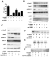
Regulation of perlecan requires autocrine TGF-β signaling, p38 MAPK and ERK signaling pathways. (a) Mechanical strain induces increased TGF-β production by human endothelial cells. Results shown are an ELISA assay after subtracting TGF-β in growth media. This effect is blocked by inhibitors of the ERK signaling pathway (U0126) and p38 MAPK signaling pathway (SB 293063). *p < 0.05 versus all other groups; **p < 0.05 significantly different from all groups except unloaded samples with SB293062; other comparisons not shown for simplicity. (b) The p38 and ERK signaling pathways are activated by 24 hrs of mechanical strain. Endothelial cells were treated with inhibitors one hour prior to exposure to 24 hrs of mechanical strain. Western blotting analysis revealed increased phosphorylation of smad 2, ERK1/2 and p38 MAPK in the absence of inhibitors. (c) Autocrine TGF-β signaling is needed for maximal phosphorylation of p38, ERK1/2 and Smad 2. Endothelial cells were treated with a neutralizing antibody to TGF-β for 1 hr prior to 24 hrs of cyclic mechanical strain. Western blotting analysis of intracellular signaling intermediates showed reduced activation of Smad 2, ERK1/2, and p38 by mechanical strain in the presence of a neutralizing antibody to TGF-β. (d) Inhibitors to p38 or MEK block load-mediated increases in soluble perlecan. (e) A neutralizing antibody to TGF-β blocks load-induced increase in soluble perlecan. (f) Addition of 100 pg/ml TGF-β increased perlecan expression even in the absence of mechanical strain. An inhibitor to TGF-β receptor I signaling, 200 nM of [3-(Pyridin-2-yl)-4-(4-quinonyl)] -1H-pyrazole, blocked perlecan upregulation by mechanical load.
Figure 5
Figure 5
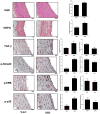
Immunohistochemical analysis of the aorta from wild-type (WKY) and spontaneously hypertensive rats (SHR) revealed increased endothelial heparan sulfate proteoglycan (HSPG) and endothelial staining for TGF-β, phospho-ERK, phospho-p38, and phospho-Smad 2. Rats were aged to 20 weeks and the aortae harvested and processed for paraffin sections (Bar = 100 μm). Staining for HSPGs was performed by deparaffinizing the sections and digesting for 2 hrs with 48 mU/ml heparitinase III. An antibody was used that recognizes the heparan sulfate “stub” following digestion with heparitinase (3G10). *p < 0.05 versus wild-type arteries (n = 8).
Figure 6
Figure 6
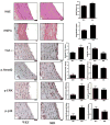
Measurement of arterial cell signaling in an ex-vivo arterial culture system. (a) Endothelial integrity is maintained in the ex-vivo culture system. Immunofluorescence staining for PECAM-1 is shown in red and staining for nuclei in blue (Bar = 200 μm). (b) Immunohistochemical analysis of ex-vivo aortae treated with inhibitors and exposed to pulsatile pressure and flow for 24 hrs. (c) In the absence of inhibitors high pressure led to an increase in endothelial HSPG, TGF-β, phospho-Smad 2 and phospho-ERK. Upregulation of endothelial HSPG, TGF-β, phospho-Smad 2 and phospho-ERK by high pressure was blocked by a neutralizing antibody to TGF-β or U0126. In this system, phospho-p38 was not significantly upregulated by high pressure and inhibition of p38 activity (SB293063) did not affect the upregulation of endothelial HSPG, TGF-β, phospho-Smad 2 and phospho-ERK by high pressure. Bar = 100 μm. *p < 0.05 versus normal pressure, control arteries.

Similar articles

Cited by

References

    1. Libby P, Aikawa M, Jain MK. Vascular endothelium and atherosclerosis. Handb Exp Pharmacol. 2006;176:285–306. - PubMed
    1. Pober JS. Warner-Lambert/Parke-Davis award lecture. Cytokine-mediated activation of vascular endothelium. Physiology and pathology. Am J Pathol. 1988;133:426–433. - PMC - PubMed
    1. Davies PF. Flow-mediated endothelial mechanotransduction. Physiol Rev. 1995;75:519–560. - PMC - PubMed
    1. Li YS, Haga JH, Chien S. Molecular basis of the effects of shear stress on vascular endothelial cells. J Biomech. 2005;38:1949–1971. - PubMed
    1. Harrison DG, Widder J, Grumbach I, Chen W, Weber M, Searles C. Endothelial mechanotransduction, nitric oxide and vascular inflammation. J Intern Med. 2006;259:351–363. - PubMed

Publication types

MeSH terms

Substances