. 2008 Jul 18;103(2):212-20.
doi: 10.1161/CIRCRESAHA.108.176230. Epub 2008 Jun 26.

Role of nox2-based NADPH oxidase in bone marrow and progenitor cell function involved in neovascularization induced by hindlimb ischemia

Affiliations

Role of nox2-based NADPH oxidase in bone marrow and progenitor cell function involved in neovascularization induced by hindlimb ischemia

Norifumi Urao et al. Circ Res. .

Abstract

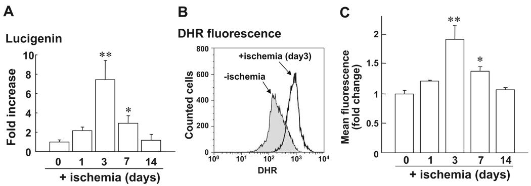
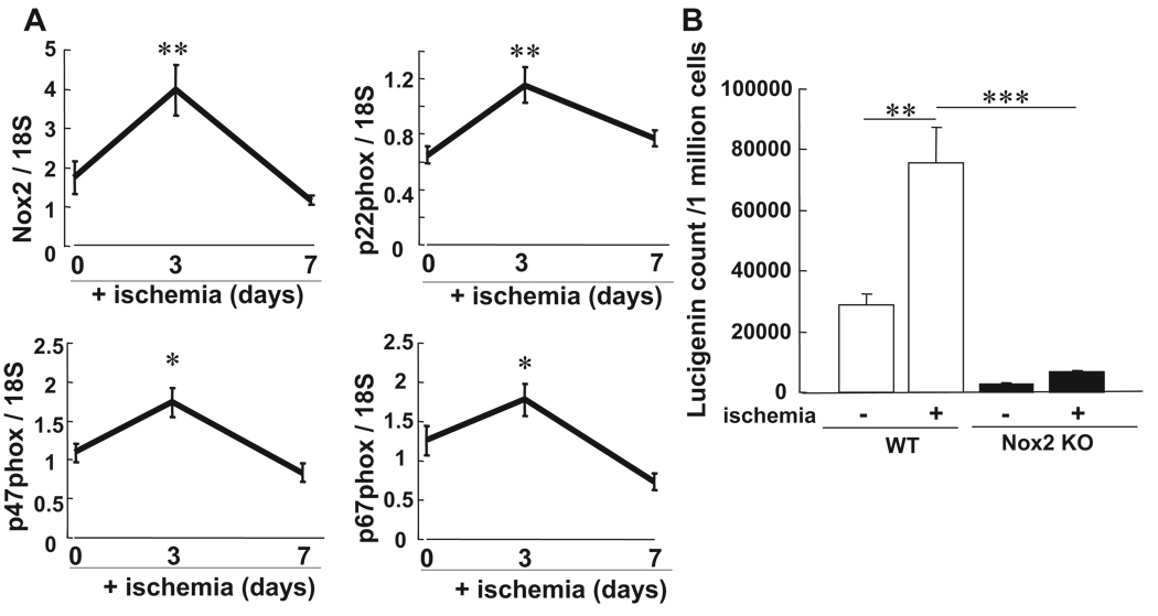
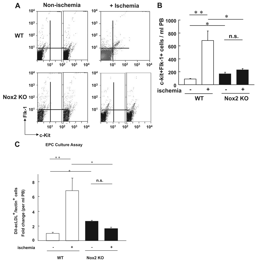
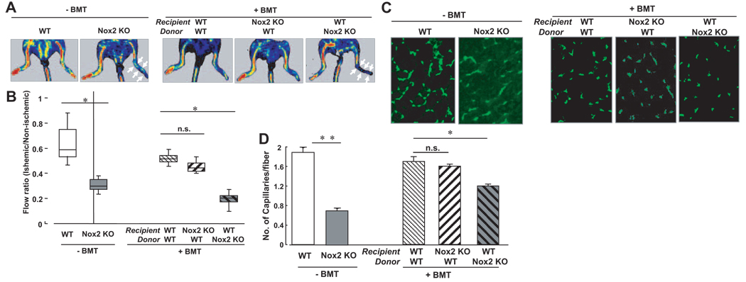
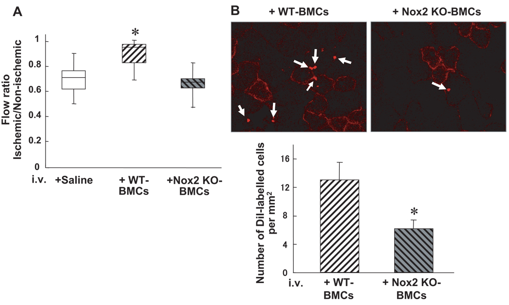
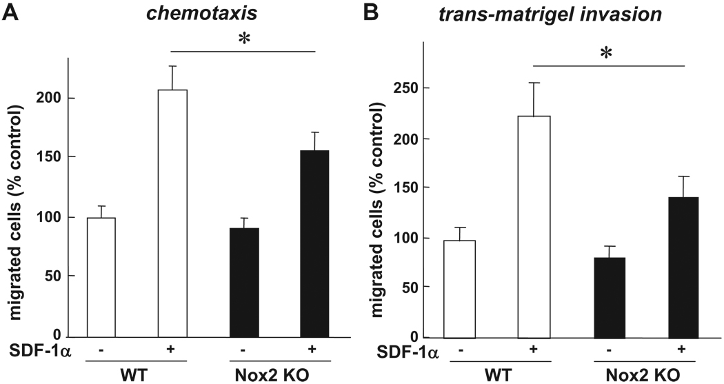
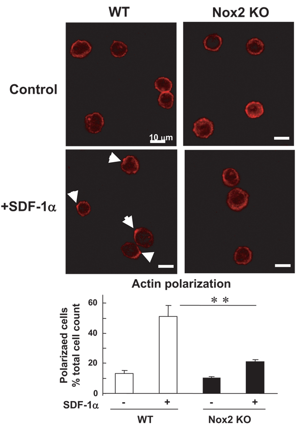
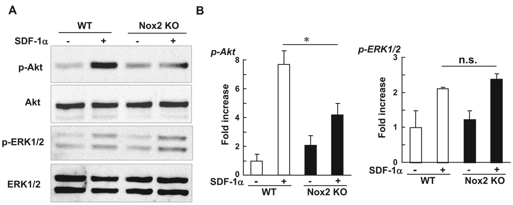
Bone marrow (BM) is the major reservoir for endothelial progenitor cells (EPCs). Postnatal neovascularization depends on not only angiogenesis but also vasculogenesis, which is mediated through mobilization of EPCs from BM and their recruitment to the ischemic sites. Reactive oxygen species (ROS) derived from Nox2-based NADPH oxidase play an important role in postnatal neovascularization; however, their role in BM and EPC function is unknown. Here we show that hindlimb ischemia of mice significantly increases Nox2 expression and ROS production in BM-mononuclear cells (BMCs), which is associated with an increase in circulating EPC-like cells. Mice lacking Nox2 show reduction of ischemia-induced flow recovery, ROS levels in BMCs, as well as EPC mobilization from BM. Transplantation of wild-type (WT)-BM into Nox2-deficient mice rescues the defective neovascularization, whereas WT mice transplanted with Nox2-deficient BM show reduced flow recovery and capillary density compared to WT-BM transplanted control. Intravenous infusion of WT- and Nox2-deficient BMCs into WT mice reveals that neovascularization and homing capacity are impaired in Nox2-deficient BMCs in vivo. In vitro, Nox2-deficient c-kit+Lin- BM stem/progenitor cells show impaired chemotaxis and invasion as well as polarization of actins in response to stromal derived factor (SDF), which is associated with blunted SDF-1-mediated phosphorylation of Akt. In conclusion, Nox2-derived ROS in BM play a critical role in mobilization, homing, and angiogenic capacity of EPCs and BM stem/progenitor cells, thereby promoting revascularization of ischemic tissue. Thus, NADPH oxidase in BM and EPCs is potential therapeutic targets for promoting neovascularization in ischemic cardiovascular diseases.


Figures

Figure 1
Figure 1
Increase of ROS production in BMCs after hindlimb ischemia. Low-density BM-MNCs (BMCs) were separated by Histopaque 1077. A, O2∙− production in BMCs after hindlimb ischemia on postoperative days 0, 1, 3, 7, and 14, as measured by lucigenin-enhanced chemiluminescence technique (n=4 in each time point). B, Dihydrorhodamine 123 (DHR) fluorescence in BMCs after hindlimb ischemia is measured by FACS analysis. Upper panel shows the representative histogram of fluorescence intensity, expressed by the logarithum (x axis) on 0 and 3 days after ischemia. Lower panel shows the averaged data, expressed as fold change of fluorescence over basal (the ratio on day 0 was set to 1) on various postoperative days (n=4, **P<0.05, **P<0.01 vs day 0).
Figure 2
Figure 2
Nox2-based NADPH oxidase is involved in ROS production in BMCs after hindlimb ischemia. A, The mRNA expression for Nox2 and its coupled NADPH oxidase components including p22phox, p47phox, and p67phox in BMCs at 0, 3, and 7 days after ischemia, as measured by real-time PCR. B, O2∙− production in BMCs at 0 and 3 days after hindlimb ischemia in WT and Nox2−/− mice, as measured by lucigenin-enhanced chemiluminescence technique (n=4 to 6, **P<0.01 vs baseline, ***P<0.001 vs WT).
Figure 3
Figure 3
The number of circulating EPC-like cells after hindlimb ischemia is inhibited in Nox2−/− mice. A, The number of EPC-like ckit+Flk1+ cells in peripheral blood (PB) mononuclear cell fraction at 0 (+ nonischemia) and 3 days (+ ischemia) after hindlimb ischemia in WT and Nox2−/− mice, as measured by FACS analysis. B, Statistical analysis of ckit+Flk1+ positive cells in PB (n=6, *P<0.05, **P<0.01). C, EPC culture assay. Quantitative analysis of the numbers of DiI-acLDL and BS lectin double positive EPCs measured at 4 days after culture of PB-derived MNCs obtained from WT and Nox2−/− mice at 0 and 3 days after ischemia. Data are expressed as fold change over basal (the ratio on day 0 in WT mice was set to 1). (n=4 to 6, *P<0.05, **P<0.01).
Figure 4
Figure 4
Transplantation of BM from wild-type mice rescues the impaired neovascularization after hindlimb ischemia in Nox2−/− mice. The lethally irradiated recipient WT or Nox2−/− mice were transplanted with BMCs from WT or Nox2−/− mice and subjected to hindlimb ischemia at 6 to 8 weeks after BM transplantation. A, Representative picture of laser Doppler blood flow (LDBF) imaging at day 14 of ischemic (right) and nonischemic (left) limbs after ligation of femoral artery in WT and Nox2−/− mice without (−) or with (+) BM transplantation (BMT). Arrow indicates delayed blood flow recovery. B, Quantification of blood flow recovery, as determined by the ischemic/nonischemic LDBF ratio in each group (n=4 to 10, *P<0.05). C, Representative pictures of immunostaining of isolectin B4 positive cells which represent capillaries in ischemic tissues obtained from WT and Nox2−/− mice without or with BMT, as described above. D, Quantitative analysis of capillary density, expressed as the number of capillaries per fiber in each group (n=4, *P<0.05, **P<0.01).
Figure 5
Figure 5
Homing and neovascularization capacity are impaired in Nox2−/− BMCs in vivo. A, BMCs (3×10⁶ cells) from WT or Nox2−/− mice, or saline control, were intravenously injected into WT mice at 24 hours after femoral artery ligation, and blood flow recovery, as assessed by the ischemic/nonischemic LDBF ratio, was measured at 14 days after hindlimb ischemia. (n=4, *P<0.05 vs control). B, Upper panel, representative photographs of accumulated intravenously injected Dil-labeled WT- and Nox2−/− BMCs in ischemic adductor muscles. Lower panel, quantitative analysis of the number of accumulated Dil-labeled WT- and Nox2−/− BMCs in ischemic border zones of adductor muscle (n=3 in each group, *P<0.05).
Figure 6
Figure 6
Chemotaxis and transmatrigel invasion induced by SDF-1α are impaired in Nox2−/− BM stem/progenitor cells in vitro. Quantification of SDF-1α–induced migration (chemotaxis; A) and invasion through matrigel (B) of WT-and Nox2−/− BM c-kit+Lin progenitor cells, as measured by transwell migration assay. Cells were stimulated with or without SDF-1α (300 ng/mL) for 3 hours. Bar graph represents averaged data, expressed as fold change in number of migrated cells to lower chamber, over that in unstimulated WT cells (control). (n=4, *P<0.05).
Figure 7
Figure 7
SDF-1α–induced F-actin polarization is impaired in Nox2−/− BM stem/progenitor cells in vitro. Upper panel, freshly isolated WT- and Nox2−/− BM c-kit+Lin cells in suspension were stimulated with or without SDF-1α for 5 minutes, fixed, and then stained for Alexa Fluor 568 conjugated phalloidin, which is visualized by confocal microscopy. Arrows indicate the polarized F-actin. Original magnification is ×630 and bars show 10 µm. Lower panel, quantitative analysis of SDF-induced F-actin polarization in WT- and Nox2−/− BM c-kit+Lin cells. Data are expressed as percentage of the total 100 cells from 4 randomly selected fields (×100; n=4, **P<0.01).
Figure 8
Figure 8
Phosphorylation of Akt induced by SDF-1 is inhibited in Nox2−/− BMCs. Freshly isolated WT- and Nox2−/− BMCs were serum depleted for 4 hours and then stimulated with SDF-1α (300 ng/mL) for 5 minutes. Lysates were used for Western analysis with antiphospho-Akt (Ser473), phospho-ERK1/2, total Akt, or total ERK1/2 antibodies. Data are expressed as fold increase over the unstimulated WT cells (n=3, *P<0.05).

References

    1. Isner JM, Losordo DW. Therapeutic angiogenesis for heart failure. Nat Med. 1999;5:491–492. - PubMed
    1. Shintani S, Murohara T, Ikeda H, Ueno T, Sasaki K, Duan J, Imaizumi T. Augmentation of postnatal neovascularization with autologous bone marrow transplantation. Circulation. 2001;103:897–903. - PubMed
    1. Morrison SJ, Shah NM, Anderson DJ. Regulatory mechanisms in stem cell biology. Cell. 1997;88:287–298. - PubMed
    1. Kiger AA, White-Cooper H, Fuller MT. Somatic support cells restrict germline stem cell self-renewal and promote differentiation. Nature. 2000;407:750–754. - PubMed
    1. Lapidot T, Petit I. Current understanding of stem cell mobilization: the roles of chemokines, proteolytic enzymes, adhesion molecules, cytokines, and stromal cells. Exper Hematol. 2002;30:973–981. - PubMed

Publication types

MeSH terms