

Atelectasis induced by thoracotomy causes lung injury during mechanical ventilation in endotoxemic rats
- PMID: 18583875
- PMCID: PMC2526521
- DOI: 10.3346/jkms.2008.23.3.406
Atelectasis induced by thoracotomy causes lung injury during mechanical ventilation in endotoxemic rats
Abstract
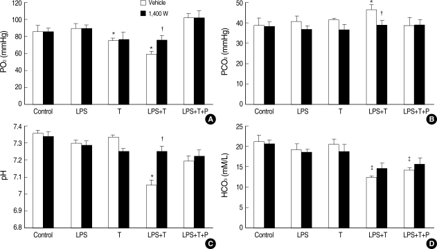
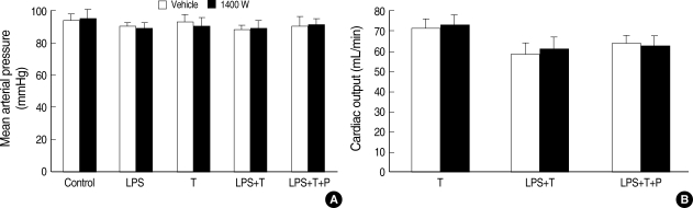
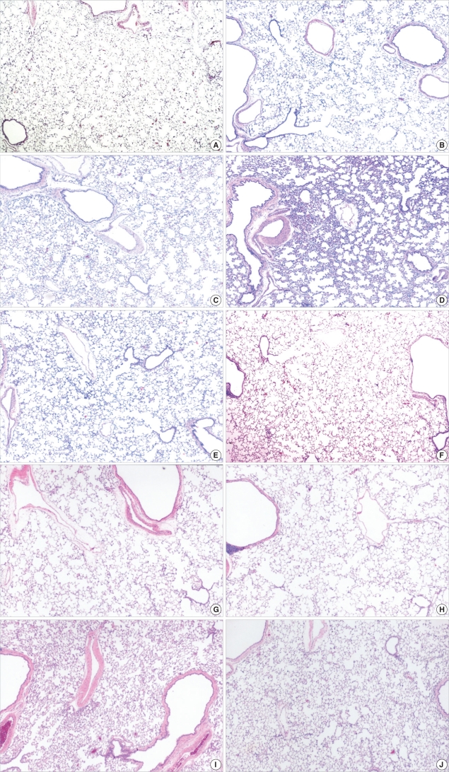
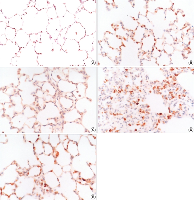
Atelectasis can impair arterial oxygenation and decrease lung compliance. However, the effects of atelectasis on endotoxemic lungs during ventilation have not been well studied. We hypothesized that ventilation at low volumes below functional residual capacity (FRC) would accentuate lung injury in lipopolysaccharide (LPS)-pretreated rats. LPS-pretreated rats were ventilated with room air at 85 breaths/min for 2 hr at a tidal volume of 10 mL/kg with or without thoracotomy. Positive end-expiratory pressure (PEEP) was applied to restore FRC in the thoracotomy group. While LPS or thoracotomy alone did not cause significant injury, the combination of endotoxemia and thoracotomy caused significant hypoxemia and hypercapnia. The injury was observed along with a marked accumulation of inflammatory cells in the interstitium of the lungs, predominantly comprising neutrophils and mononuclear cells. Immunohistochemistry showed increased inducible nitric oxide synthase (iNOS) expression in mononuclear cells accumulated in the interstitium in the injury group. Pretreatment with PEEP or an iNOS inhibitor (1400 W) attenuated hypoxemia, hypercapnia, and the accumulation of inflammatory cells in the lung. In conclusion, the data suggest that atelectasis induced by thoracotomy causes lung injury during mechanical ventilation in endotoxemic rats through iNOS expression.
Figures
Similar articles
-
Volume recruitment maneuvers are less deleterious than persistent low lung volumes in the atelectasis-prone rabbit lung during high-frequency oscillation.Crit Care Med. 1993 Mar;21(3):402-12. doi: 10.1097/00003246-199303000-00019. Crit Care Med. 1993. PMID: 8440111
-
Role of tidal volume, FRC, and end-inspiratory volume in the development of pulmonary edema following mechanical ventilation.Am Rev Respir Dis. 1993 Nov;148(5):1194-203. doi: 10.1164/ajrccm/148.5.1194. Am Rev Respir Dis. 1993. PMID: 8239153
-
Protective effects of dexmedetomidine-ketamine combination against ventilator-induced lung injury in endotoxemia rats.J Surg Res. 2011 May 15;167(2):e273-81. doi: 10.1016/j.jss.2010.02.020. Epub 2010 Mar 12. J Surg Res. 2011. PMID: 20452617
-
Review: artifical ventilation with positive end-expiratory pressure (PEEP). Historical background, terminology and patho-physiology.Eur J Intensive Care Med. 1976 Sep;2(2):77-85. doi: 10.1007/BF01886120. Eur J Intensive Care Med. 1976. PMID: 786640 Review.
-
Atelectasis formation during anesthesia: causes and measures to prevent it.J Clin Monit Comput. 2000;16(5-6):329-35. doi: 10.1023/a:1011491231934. J Clin Monit Comput. 2000. PMID: 12580216 Review.
Cited by
-
Positive end-expiratory pressure attenuates positional effect after thoracotomy.Ann Thorac Med. 2014 Apr;9(2):112-9. doi: 10.4103/1817-1737.128860. Ann Thorac Med. 2014. PMID: 24791175 Free PMC article.
References
-
- Rehder K, Sessler AD, Marsh HM. General anesthesia and the lung. Am Rev Respir Dis. 1975;112:541–563. - PubMed
-
- Lindberg P, Gunnarsson L, Tokics L, Secher E, Lundquist H, Brismar B, Hedenstierna G. Atelectasis and lung function in the postoperative period. Acta Anaesthesiol Scand. 1992;36:546–553. - PubMed
-
- Reissmann H, Bohm SH, Suarez-Sipmann F, Tusman G, Buschmann C, Maisch S, Pesch T, Thamm O, Plumers C, Schulte am Esch J, Hedenstierna G. Suctioning through a double-lumen endotracheal tube helps to prevent alveolar collapse and to preserve ventilation. Intensive Care Med. 2005;31:431–440. - PubMed
-
- Muller JM, Erasmi H, Stelzner M, Zieren U, Pichlmaier H. Surgical therapy of oesophageal carcinoma. Br J Surg. 1990;77:845–857. - PubMed
-
- Tandon S, Batchelor A, Bullock R, Gascoigne A, Griffin M, Hayes N, Hing J, Shaw I, Warnell I, Baudouin SV. Peri-operative risk factors for acute lung injury after elective oesophagectomy. Br J Anaesth. 2001;86:633–638. - PubMed
Publication types
MeSH terms
Substances
Grants and funding
LinkOut - more resources
Full Text Sources
Medical
