

New roles for the major human 3'-5' exonuclease TREX1 in human disease
- PMID: 18583934
- PMCID: PMC2825026
- DOI: 10.4161/cc.7.12.6162
New roles for the major human 3'-5' exonuclease TREX1 in human disease
Abstract
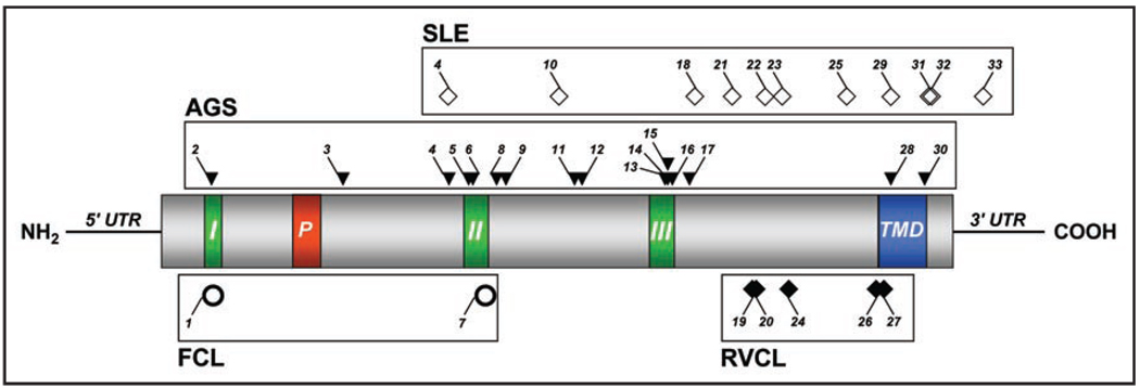
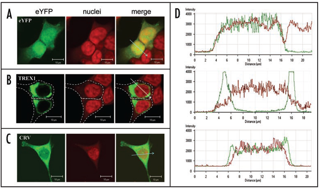
Aicardi-Goutières syndrome (AGS), Systemic Lupus Erythematosus (SLE), Familial Chilblain Lupus (FCL) and Retinal Vasculopathy and Cerebral Leukodystrophy (RVCL) {a new term encompassing three independently described conditions with a common etiology--Cerebroretinal Vasculopathy (CRV), Hereditary Vascular Retinopathy (HVR) and Hereditary Endotheliopathy, Retinopathy and Nephropathy (HERNS)}--have previously been regarded as distinct entities. However, recent genetic analysis has demonstrated that each of these diseases maps to chromosome 3p21 and can be caused by mutations in TREX1, the major human 3'-5' exonuclease. In this review, we discuss the putative functions of TREX1 in relationship to the clinical, genetic and functional characteristics of each of these conditions.
Figures
References
-
- Richards A, van den Maagdenberg AM, Jen JC, et al. Truncations in the carboxyl-terminus of human 3'–5' DNA exonuclear TREX1 cause autosomal dominant retinal vasculopathy with cerebral leukodystrophy. Nat Genet. 2007;39:1068–1070. - PubMed
-
- Crow YJ, Hayward BE, Parmar R, et al. Mutations in the gene encoding the 3’–5’ DNA exonuclease TREX1 cause Aicardi-Goutieres syndrome at the AGS1 locus. Nat Genet. 2006;38:917–920. - PubMed
Publication types
MeSH terms
Substances
Grants and funding
LinkOut - more resources
Full Text Sources
Other Literature Sources
Medical
Molecular Biology Databases