. 2008 Aug 6;27(15):2135-46.
doi: 10.1038/emboj.2008.126. Epub 2008 Jun 26.

The DEAD-box helicase DDX3X is a critical component of the TANK-binding kinase 1-dependent innate immune response

Affiliations

The DEAD-box helicase DDX3X is a critical component of the TANK-binding kinase 1-dependent innate immune response

Didier Soulat et al. EMBO J. .

Abstract

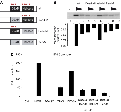
TANK-binding kinase 1 (TBK1) is of central importance for the induction of type-I interferon (IFN) in response to pathogens. We identified the DEAD-box helicase DDX3X as an interaction partner of TBK1. TBK1 and DDX3X acted synergistically in their ability to stimulate the IFN promoter, whereas RNAi-mediated reduction of DDX3X expression led to an impairment of IFN production. Chromatin immunoprecipitation indicated that DDX3X is recruited to the IFN promoter upon infection with Listeria monocytogenes, suggesting a transcriptional mechanism of action. DDX3X was found to be a TBK1 substrate in vitro and in vivo. Phosphorylation-deficient mutants of DDX3X failed to synergize with TBK1 in their ability to stimulate the IFN promoter. Overall, our data imply that DDX3X is a critical effector of TBK1 that is necessary for type I IFN induction.


Figures

Figure 1
Figure 1
The DEAD-box helicase DDX3X is a target of TBK1. (A) Schematic representation of the tandem affinity purification protocol: GS-TAP-tagged TBK1 expressed in RAW264.7 cells was purified using rabbit immunoglobulin G (IgG) agarose and eluted by tobacco etch virus (TEV) protease cleavage. Next, the remaining complex was purified using streptavidin agarose and eluted by boiling in SDS sample buffer. (B) The final TAP eluate was separated on an SDS–PAGE and stained by silver staining. Protein complex composition was analysed by LC-MSMS. The position of several complex components is depicted next to the region where it was identified.
Figure 2
Figure 2
DDX3X is required for IFN-β induction. Expression of TBK1 or DDX3X was targeted in RAW264.7 macrophages using specific siRNAs. Expression levels of TBK1 (A) or DDX3X (B) were monitored by immunoblotting. Pan-ERK was monitored as a loading control. (C) siRNA-treated RAW264.7 cells were infected with L. monocytogenes for 4 h. Induction of IFN-β mRNA (left panel), RANTES mRNA (middle panel) and TNF-α mRNA (right panel) were measured by quantitative RT–PCR. (D) Similar cells were transfected with poly(I:C) (left panel), treated with LPS (middle panel) or transfected with poly(dA:dT) (right panel). Induction of IFN-β mRNA was measured by quantitative RT–PCR. (E) siRNA-treated RAW264.7 cells were infected with L. monocytogenes or treated with IFN-β for 4 h. Induction of IRF7 and Mx2 was measured by quantitative RT–PCR. Sets of data were analysed using the paired Student's t-test (two-tailed, equal variance). Statistical significance was assessed based on the P-value: *P<0.05, **P<0.01 and ***P<0.001.
Figure 3
Figure 3
TBK1 has an impact on Rev/DDX3X-dependent nuclear RNA export. (A) Schematic representation of the Rev reporter construct pDM128. In the absence of Rev-mediated nuclear mRNA export, the CAT gene is spliced and therefore not translated. If Rev is present, the mRNA is exported out of the nucleus, splicing is prevented and the CAT mRNA is translated. (B) HEK293 cells were transfected with the reporter plasmid pDM128 and constant amounts of Rev DDX3X wt or K230A and TBK1 wt or K38M were added as indicated. CAT expression was measured 24 h post-transfection by ELISA (Roche).
Figure 4
Figure 4
DDX3X and TBK1 led to synergistic activation of the IFN-β promoter. Cells were transfected with a firefly luciferase reporter plasmid and a Renilla luciferase plasmid. Firefly luciferase activity was measured after 24 h and normalized to Renilla luciferase activity. (A) HEK293 cells were transfected with the IFN-β reporter plasmid and 1 μg of plasmids encoding MAVS, DDX3X or DDX3X-K230A and 500 ng of a TBK1 expression plasmid as indicated. (B) HEK293 cells were transfected with the NF-κB reporter plasmid and 1 μg of plasmids encoding HA-tagged MAVS, DDX3X or DDX3X-K230A and 500 ng of a TBK1 expression plasmid as indicated. (C) MEFs from wt mice (black bars), TBK1-deficient mice (grey bars) or IRF3-deficient mice (white bars) were transfected with the IFN-β reporter plasmid and 1 μg of plasmids encoding HA-tagged MAVS, DDX3X, TBK1 and TBK1-S172A as indicated.
Figure 5
Figure 5
DDX3X is recruited to the enhanceosome-binding site on the IFN-β promoter. (A) Schematic representation of the IFN-β promoter. Enhanceosome-binding site (white box) is flanked by specific primers, whereas a 1 kb upstream region is flanked by control primers. (B) RAW264.7 macrophages were infected with L. monocytogenes for 0–3 h. Quantitative PCR was realized on ChIP samples treated with either an IRF3-specific antiserum (black bar) or an unrelated serum (white bar). (C) RAW264.7 NTAP(GS)-DDX3X was infected with L. monocytogenes for 0–3 h. Quantitative PCR was realized on samples immunoprecipitated with IgG beads with primers specific for either the enhanceosome-binding site (black bar) or the control region (white bar). Sets of data were analysed using the paired Student's t-test (two-tailed, equal variance). Statistical significance was assessed based on the P-value: *P<0.05 and **P<0.01.
Figure 6
Figure 6
DDX3X is phosphorylated by TBK1. (A, B) HEK293 cells were transiently transfected with Myc- or HA-tagged constructs as indicated. Cells were treated with 10 μM staurosporine 1 h before lysis as indicated. Cells were lysed 48 h post transfection. Cell extracts were treated with 5 U CIP for 1 h as indicated. Cell extracts were analysed by immunoblotting using either anti-Myc (Rockland), anti-HA (Covance Research) or an antiserum against phosphoserine 172 in TBK1. (C) GST-tagged IRF3, DDX3X and Grb2 were expressed in E. coli and purified by HisTrap chromatography. Purity was assessed by Coomassie staining of three different dilutions of each protein (right panel). TBK1-mediated phosphorylation was assessed by incubating the three potential substrates (IRF3, DDX3X and Grb2) with TBK1 in the presence of [γ-32P]ATP. Lane 1 contains TBK1 without substrate, whereas lanes 2, 6 and 10 contain the substrates without TBK1 (left panel).
Figure 7
Figure 7
Mapping of the TBK1 phosphorylation site in DDX3X. (A) Peptides derived from DDX3X or control peptides derived from TBK1 or IRF3 along with alanine mutants were incubated with TBK1 in the presence of [γ-32P]ATP. Each array contained a total of 82 peptides spotted in triplicate. (B) Phosphorylation sites obtained from the peptide array (see also text) were used to build a TBK1 phosphorylation consensus sequence. (C) Putative DDX3X phosphorylation sites were mapped onto the structure of a truncated version of DDX3X.
Figure 8
Figure 8
Phosphorylation-deficient DDX3X mutants fail to synergize with TBK1. (A) Phosphorylation sites obtained from the peptide array were mutated as follows: Dead-M (S181A, S183A, S240A and S269A), Helic-M (S429A, T438A, S442A, S456A and S520A) or Pan-M (S181A, S183A, S240A, S269A, S429A, T438A, S442A, S456A and S520A). (B) Mutated DDX3X proteins were purified from E. coli and analysed in the in vitro kinase assay as described in Figure 6B. (C) HEK293 cells were transiently transfected with the IFN-β reporter plasmid and MAVS, DDX3X wt, different DDX3X mutants (Dead-M, Helic-M and Pan-M) and TBK1 as indicated. Reporter activity was quantified as described in Figure 4.

Similar articles

Cited by

References

    1. Akira S (2006) TLR signaling. Curr Top Microbiol Immunol 311: 1–16 - PubMed
    1. Ariumi Y, Kuroki M, Abe K, Dansako H, Ikeda M, Wakita T, Kato N (2007) DDX3 DEAD-box RNA helicase is required for hepatitis C virus RNA replication. J Virol 81: 13922–13926 - PMC - PubMed
    1. Botlagunta M, Vesuna F, Mironchik Y, Raman A, Lisok A, Winnard P Jr, Mukadam S, Van Diest P, Chen JH, Farabaugh P, Patel AH, Raman V (2008) Oncogenic role of DDX3 in breast cancer biogenesis. Oncogene; doi:10.1038/onc.2008.33 - PMC - PubMed
    1. Bouwmeester T, Bauch A, Ruffner H, Angrand PO, Bergamini G, Croughton K, Cruciat C, Eberhard D, Gagneur J, Ghidelli S, Hopf C, Huhse B, Mangano R, Michon AM, Schirle M, Schlegl J, Schwab M, Stein MA, Bauer A, Casari G et al. (2004) A physical and functional map of the human TNF-alpha/NF-kappa B signal transduction pathway. Nat Cell Biol 6: 97–105 - PubMed
    1. Burckstummer T, Bennett KL, Preradovic A, Schutze G, Hantschel O, Superti-Furga G, Bauch A (2006) An efficient tandem affinity purification procedure for interaction proteomics in mammalian cells. Nat Methods 3: 1013–1019 - PubMed

Publication types

MeSH terms