. 2008 Aug;9(8):802-9.
doi: 10.1038/embor.2008.90. Epub 2008 Jun 27.

The nature and character of the transition state for the ADP-ribosyltransferase reaction

Affiliations

The nature and character of the transition state for the ADP-ribosyltransferase reaction

René Jørgensen et al. EMBO Rep. 2008 Aug.

Abstract

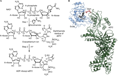
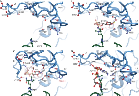
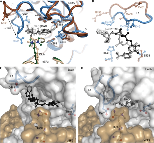
Exotoxin A (ExoA) from Pseudomonas aeruginosa is an important virulence factor that belongs to a class of exotoxins that are secreted by pathogenic bacteria which cause human diseases such as cholera, diphtheria, pneumonia and whooping cough. We present the first crystal structures, to our knowledge, of ExoA in complex with elongation factor 2 (eEF2) and intact NAD(+), which indicate a direct role of two active-site loops in ExoA during the catalytic cycle. One loop moves to form a solvent cover for the active site of the enzyme and reaches towards the target residue (diphthamide) in eEF2 forming an important hydrogen bond. The NAD(+) substrate adopts a conformation remarkably different from that of the NAD(+) analogue, betaTAD, observed in previous structures, and fails to trigger any loop movements. Mutational studies of the two loops in the toxin identify several residues important for catalytic activity, in particular Glu 546 and Arg 551, clearly supporting the new complex structures. On the basis of these data, we propose a transition-state model for the toxin-catalysed reaction.


Conflict of interest statement

The authors declare that they have no conflict of interest.

Figures

Figure 1
Figure 1
Reaction mechanism and structure of ExoA. (A) Schematic representation of the reaction catalysed by ExoA. ExoA catalyses the transfer of A-ribose from NAD+ to N3 of the diphthamide imidazole, impairing eEF2 function and causing inhibition of protein synthesis. (B) Ribbon representation of the eEF2–ExoAc–NAD+ complex. The ExoAc (blue) is bound to the diphthamide-containing domain of eEF2 (green) with the diphthamide (Diph; red ball and stick) pointing towards the NAD+ (black ball and stick). A-ribose, ADP ribose; A-phosphate, adenine phosphate; eEF2, elongation factor 2; ExoA, exotoxin A; N-ribose, nicotinamide ribose.
Figure 2
Figure 2
Sequence of events for the ADP-ribosylation of eEF2. (A) The structure of the eEF2–ExoAc complex before NAD+ binding. ExoAc is shown in blue, eEF2 in green and hydrogen bonds in purple. Residues surrounding the NAD+ binding pocket are shown as a ball-and-stick representation (Protein Data Bank accession code 1ZM3; Jorgensen et al, 2005). (B) Structure of the eEF2–ExoAc–βTAD complex with the trimethyl ammonium group of the diphthamide (Diph; green) reaching towards the nicotinamide phosphate of βTAD (black carbons; Protein Data Bank accession code 1ZM4; Jorgensen et al, 2005). (C) The structure of the eEF2–ExoAc(R551H)–NAD+ complex with L1 hydrogen bonding to the NAD+ (grey carbons) and the diphthamide. Several water molecules (brown spheres) mediate contact between NAD+ and ExoAc. (D) The ADP-ribosyltransferase reaction product, the ADPR–eEF2–ExoAc complex (Protein Data Bank accession code 1ZM2; Jorgensen et al, 2005). The ADP-ribose (grey carbons) has been transferred from NAD+ (donor) to N3 of the imidazole ring of the diphthamide residue. The nicotinamide is docked into the binding pocket. ExoA, exotoxin A; eEF2, elongation factor 2; L, loop.
Figure 3
Figure 3
The NAD+ binding pocket. (A) A comparison of the NAD+-binding site in the eEF2–ExoAc–βTAD complex and the eEF2–ExoAc(R551H)–NAD+ mutant complex. In the eEF2–ExoAc–βTAD complex, ExoAc is shown in dark brown, eEF2 in light brown and βTAD in black. In the eEF2–ExoAc(R551H)–NAD+ complex, ExoAc is shown in blue, eEF2 in dark green and NAD+ in grey. Residues involved in coordinating NAD+ are shown as a ball-and-stick representation. (B) Conformation of L1, and the NAD+ and βTAD legends in the NAD+-binding site as viewed from the diphthamide residue. When compared with (A), the view is rotated −90° around the x axis and 90° around the z axis. Residue and legend colours are as shown in (A). (C) Transparent surface representation of the complex. Open conformation of L1 (blue cartoon with Asp 461 (D 461) in a ball-and-stick representation) in the eEF2–ExoAc–βTAD complex. The βTAD (black), the diphthamide (white carbons) and the ExoAc E551H (blue) are shown as a ball-and-stick representation. (D) Closed conformation of L1 in the eEF2–ExoAc(E551H)–NAD+ complex. The colouring is as in (C), except for NAD+ (grey ball and stick). ExoA, exotoxin A; eEF2, elongation factor 2; L, loop.
Figure 4
Figure 4
Transition-state model for the ADP-ribosyltransferase reaction. (A) The imidazole ring of the diphthamide residue (Diph; dark green; N3 nucleophile, cyan) migrates towards the nicotinamide ribose as the TMA group of the diphthamide moves between L1 of the toxin and the NAD+ phosphates. The original diphthamide position from the eEF2–ExoAc(R551H)–NAD+ structure is shown in light brown with Gly 701, Gly 702 and Gly 703 in red. Active-site residues of ExoAc are shown as light blue ball and sticks, with the position of Glu 546 (before roamer rotation) and Arg 551 shown in brown. ADP-ribose and nicotinamide carbons are shown in grey (C1 electrophile, dark grey). Known hydrogen bonds are shown in purple, whereas potential interactions during transition-state formation are indicated in yellow. (B) The electrostatic molecular surface of the binding pocket of the eEF2–ExoAc(R551H)–NAD+ complex. Charges were assigned to ExoAc during electrostatic calculations and the position of NAD+ (grey) is shown as a ball-and-stick representation. Asp 461 and Glu 546 (blue), and the diphthamide (white) are shown as a ball-and-stick representation. ExoA, exotoxin A; eEF2, elongation factor 2; L, loop; TMA, trimethyl ammonium.

References

    1. Armstrong S, Merrill AR (2001) Application of a fluorometric assay for characterization of the catalytic competency of a domain III fragment of Pseudomonas aeruginosa exotoxin A. Anal Biochem 292: 26–33 - PubMed
    1. Armstrong S, Yates SP, Merrill AR (2002) Insight into the catalytic mechanism of Pseudomonas aeruginosa exotoxin A. Studies of toxin interaction with eukaryotic elongation factor-2. J Biol Chem 277: 46669–46675 - PubMed
    1. Beattie BK, Prentice GA, Merrill AR (1996) Investigation into the catalytic role for the tryptophan residues within domain III of Pseudomonas aeruginosa exotoxin A. Biochemistry 35: 15134–15142 - PubMed
    1. Bell CE, Eisenberg D (1996) Crystal structure of diphtheria toxin bound to nicotinamide adenine dinucleotide. Biochemistry 35: 1137–1149 - PubMed
    1. Corda D, Di Girolamo M (2003) Functional aspects of protein mono-ADP-ribosylation. EMBO J 22: 1953–1958 - PMC - PubMed

Publication types

MeSH terms