Comparative Study
. 2008 Jun 27;4(6):e1000110.
doi: 10.1371/journal.pgen.1000110.

Alternative-NHEJ is a mechanistically distinct pathway of mammalian chromosome break repair

Affiliations
Comparative Study

Alternative-NHEJ is a mechanistically distinct pathway of mammalian chromosome break repair

Nicole Bennardo et al. PLoS Genet. .

Abstract

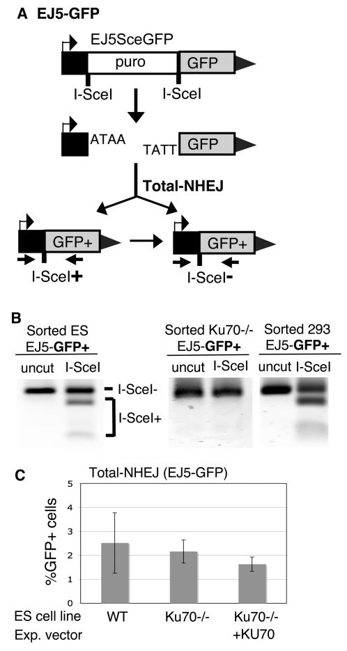
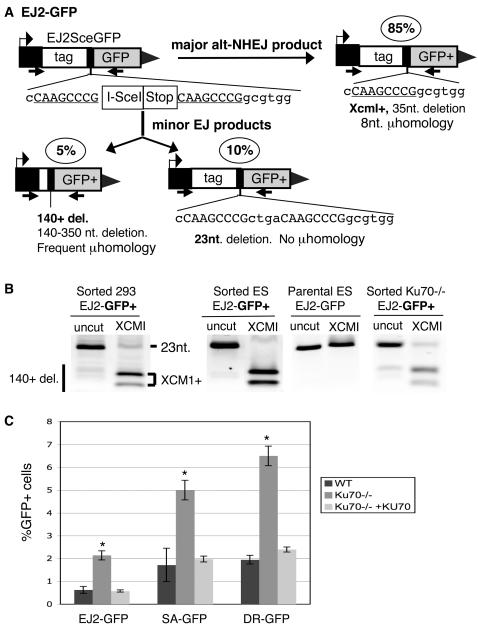
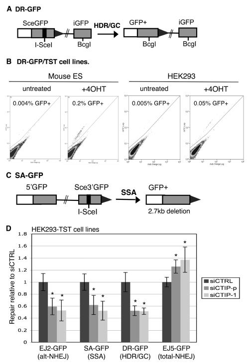
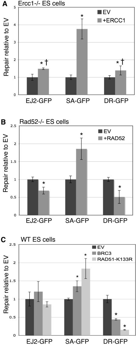
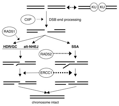
Characterizing the functional overlap and mutagenic potential of different pathways of chromosomal double-strand break (DSB) repair is important to understand how mutations arise during cancer development and treatment. To this end, we have compared the role of individual factors in three different pathways of mammalian DSB repair: alternative-nonhomologous end joining (alt-NHEJ), single-strand annealing (SSA), and homology directed repair (HDR/GC). Considering early steps of repair, we found that the DSB end-processing factors KU and CtIP affect all three pathways similarly, in that repair is suppressed by KU and promoted by CtIP. In contrast, both KU and CtIP appear dispensable for the absolute level of total-NHEJ between two tandem I-SceI-induced DSBs. During later steps of repair, we find that while the annealing and processing factors RAD52 and ERCC1 are important to promote SSA, both HDR/GC and alt-NHEJ are significantly less dependent upon these factors. As well, while disruption of RAD51 causes a decrease in HDR/GC and an increase in SSA, inhibition of this factor did not affect alt-NHEJ. These results suggest that the regulation of DSB end-processing via KU/CtIP is a common step during alt-NHEJ, SSA, and HDR/GC. However, at later steps of repair, alt-NHEJ is a mechanistically distinct pathway of DSB repair, and thus may play a unique role in mutagenesis during cancer development and therapy.


Conflict of interest statement

The authors have declared that no competing interests exist.

Figures

Figure 1
Figure 1. Total-NHEJ repair between two tandem I-SceI sites results in a variety of products.
(A) EJ5-GFP is shown along with two classes of NHEJ repair products that can restore a GFP expression cassette: one that restores an I-SceI site (I-SceI+), and one that is I-SceI–resistant (I-SceI-). (B) Restoration of the I-SceI site is common in wild-type cells, but undetectable in KU-deficient cells. EJ5-GFP was integrated into HEK293, wild-type ES, and Ku70-/- ES cells. Following transient I-SceI expression in each of these cell lines, GFP+ cells were sorted, and the GFP genes were amplified from these samples using primers depicted in (A). Shown are these products digested with I-SceI or left uncut. (C) The overall frequency of total-NHEJ is unaffected by KU deficiency. Shown is the frequency of repair of EJ5-GFP resulting in GFP+ cells from wild-type and Ku70-/- ES cells transfected in parallel with an I-SceI expression vector. Also shown are Ku70-/- ES cells cotransfected with both I-SceI and KU70 expression vectors.
Figure 2
Figure 2. Alt-NHEJ is suppressed by KU.
(A) EJ2-GFP is shown with 3 NHEJ products that are found to result in GFP+ cells. Shown in the ovals are the relative contributions of these products, based on the analysis shown in (B). The predominant GFP+ product, labeled Xcm1+, uses 8 nts of homology flanking the I-SceI site to generate an XCM1 site, resulting in a 35 nt deletion. (B) Analysis of EJ2-GFP repair products that restore the GFP+ gene. EJ2-GFP was integrated into HEK293, wild-type ES, and Ku70-/- ES cells. Using the primers shown in (A), the GFP genes were amplified from the parental ES EJ2-GFP cell line, and also from sorted GFP+ cells from each of the above cell lines following transient I-SceI expression. Shown are these amplification products, which were either uncut or cut with XCM1. (C) Repair by alt-NHEJ (EJ2-GFP) is suppressed by KU. Shown are the frequencies of alt-NHEJ repair, following transient I-SceI expression, for the wild-type and Ku70-/- EJ2-GFP cell lines, along with the Ku70-/- line co-transfected with an expression vector for KU70. Also shown are parallel experiments with the SA-GFP and DR-GFP reporters (see Figure 3). Asterisks denote a statistical difference in repair efficiency between Ku70-/- versus both wild-type, as well as Ku70-/- with transient expression of KU70 (p<0.0005).
Figure 3
Figure 3. An inducible system for I-SceI in stable cell lines used to show that siRNA-mediated disruption of CtIP affects multiple repair pathways.
(A) Shown is the structure of the DR-GFP reporter along with the HDR/GC repair product that results in GFP+ cells, as described previously in ES cells . (B) System for inducible control of I-SceI in stable cell lines. Cell lines were established with ES cells and HEK293 cells that contain the DR-GFP reporter and stable expression of the TAM-I-SceI-TAM (TST) fusion protein. These cell lines were either left untreated, or treated with 4-hydroxytamoxifen (4OHT) for a limited time (8 h for ES, 24 h for HEK293), and analyzed 3 d after starting the treatment. Shown are flow cytometric (FACS) profiles of 105 cells, where green fluorescence is plotted on the y-axis and auto orange fluorescence is on the x-axis. (C) Shown is the structure of SA-GFP reporter along with the GFP+ product of SSA repair. As discussed previously, HDR/GC associated with crossing over does not likely contribute significantly to this assay . (D) CtIP promotes alt-NHEJ, SSA and HDR, but is dispensable for total-NHEJ. HEK293 cell lines with individual reporters were exposed to control siRNA (siCTRL), a pool of three CtIP-targeting siRNAs (siCTIP-p), or a distinct single CtIP-targeting siRNA (siCTIP-1). Subsequently, I-SceI was activated by 4OHT, and repair was measured as in (B). Shown are repair frequencies relative to the mean value of siCTRL samples treated in parallel. Asterisks denote a statistical difference from siCTRL with the substrates EJ2-GFP, SA-GFP, DR-GFP, and EJ5-GFP for both siCTIP-p (p<0.0001, p = 0.0012, p<0.0001, and p = 0.0009, respectively) and siCTIP-1 (p = 0.0021, p = 0.0002, p<0.0001, and p = 0.0023, respectively).
Figure 4
Figure 4. The roles of ERCC1, RAD52, and RAD51 during alt-NHEJ, HDR/GC, and SSA.
(A) While ERCC1 significantly promotes SSA, it plays a minor role in HDR/GC and alt-NHEJ. Ercc1-/- ES cell lines with EJ2-GFP, SA-GFP, and DR-GFP were transfected with an I-SceI expression vector, along with either an expression vector for ERCC1 or empty vector (EV). Shown are the levels of repair relative to the mean value of a parallel set of EV transfections, which allows a direct comparison of the effect of complementation on the different reporters. Asterisks denote a statistical difference in repair relative to EV (alt-NHEJ and SSA, p<0.0001; DR-GFP, p = 0.0066), and the dagger denotes a statistical difference in the level of complementation relative to SA-GFP (p<0.0001). (B) RAD52 promotes SSA but not HDR/GC or alt-NHEJ. Rad52-/- ES cell lines with the reporters shown in (A) were transfected with an I-SceI expression vector, along with either an expression vector for RAD52 or empty vector. Shown are levels of repair as described in (A). Asterisks denote a statistical difference in repair relative to EV (alt-NHEJ, p = 0.0003; SA-GFP and DR-GFP, p<0.0001). (C) RAD51 promotes HDR/GC, inhibits SSA, and plays no clear role in alt-NHEJ. Wild-type ES cell lines with each of the reporters were cotransfected with an I-SceI expression vector along with either an expression vector for a BRC3 peptide derived from BRCA2, an expression vector for RAD51-K133R, or EV . Shown are levels of repair calculated relative to EV as in (A). Asterisks denote a statistical difference in repair relative to EV (SSA, p<0.016; DR-GFP, p<0.0008).
Figure 5
Figure 5. Model for the mechanistic relationships between alt-NHEJ, SSA, and HDR/GC.
Individual genetic factors, shown in ovals, are placed in the pathways based on the genetic analysis presented here, and other studies discussed in the text. End processing steps are shown as 5′ to 3′ resection, which need not be the precise mechanism in mammalian cells. The lengths of homologous annealing and 3′ end cleavage are modeled as being less extensive for both alt-NHEJ and HDR/GC relative to SSA.

References

    1. Khanna KK, Jackson SP. DNA double-strand breaks: signaling, repair and the cancer connection. Nat Genet. 2001;27:247–254. - PubMed
    1. Bassing CH, Alt FW. The cellular response to general and programmed DNA double strand breaks. DNA Repair (Amst) 2004;3:781–796. - PubMed
    1. Burma S, Chen BP, Chen DJ. Role of non-homologous end joining (NHEJ) in maintaining genomic integrity. DNA Repair (Amst) 2006;5:1042–1048. - PubMed
    1. Guirouilh-Barbat J, Huck S, Bertrand P, Pirzio L, Desmaze C, et al. Impact of the KU80 pathway on NHEJ-induced genome rearrangements in mammalian cells. Mol Cell. 2004;14:611–623. - PubMed
    1. Zhu C, Mills KD, Ferguson DO, Lee C, Manis J, et al. Unrepaired DNA breaks in p53-deficient cells lead to oncogenic gene amplification subsequent to translocations. Cell. 2002;109:811–821. - PubMed

Publication types

MeSH terms