. 2008 Sep-Oct;14(9-10):567-74.
doi: 10.2119/2008-00079.Parrish.

Modulation of TNF release by choline requires alpha7 subunit nicotinic acetylcholine receptor-mediated signaling

Affiliations

Modulation of TNF release by choline requires alpha7 subunit nicotinic acetylcholine receptor-mediated signaling

William R Parrish et al. Mol Med. 2008 Sep-Oct.

Abstract

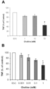
The alpha7 subunit-containing nicotinic acetylcholine receptor (alpha7nAChR) is an essential component in the vagus nerve-based cholinergic anti-inflammatory pathway that regulates the levels of TNF, high mobility group box 1 (HMGB1), and other cytokines during inflammation. Choline is an essential nutrient, a cell membrane constituent, a precursor in the biosynthesis of acetylcholine, and a selective natural alpha7nAChR agonist. Here, we studied the anti-inflammatory potential of choline in murine endotoxemia and sepsis, and the role of the alpha7nAChR in mediating the suppressive effect of choline on TNF release. Choline (0.1-50 mM) dose-dependently suppressed TNF release from endotoxin-activated RAW macrophage-like cells, and this effect was associated with significant inhibition of NF-kappaB activation. Choline (50 mg/kg, intraperitoneally [i.p.]) treatment prior to endotoxin administration in mice significantly reduced systemic TNF levels. In contrast to its TNF suppressive effect in wild type mice, choline (50 mg/kg, i.p.) failed to inhibit systemic TNF levels in alpha7nAChR knockout mice during endotoxemia. Choline also failed to suppress TNF release from endotoxin-activated peritoneal macrophages isolated from alpha7nAChR knockout mice. Choline treatment prior to endotoxin resulted in a significantly improved survival rate as compared with saline-treated endotoxemic controls. Choline also suppressed HMGB1 release in vitro and in vivo, and choline treatment initiated 24 h after cecal ligation and puncture (CLP)-induced polymicrobial sepsis significantly improved survival in mice. In addition, choline suppressed TNF release from endotoxin-activated human whole blood and macrophages. Collectively, these data characterize the anti-inflammatory efficacy of choline and demonstrate that the modulation of TNF release by choline requires alpha7nAChR-mediated signaling.


Figures

Figure 1
Figure 1
Choline inhibits TNF release (A) and NF-κB activation (B) in endotoxin-stimulated RAW-264.7 mouse macrophage-like cells. (A) RAW cells were exposed to the indicated concentration of choline 10 min prior to the addition of endotoxin (4 ng/mL). Culture supernatants were harvested 4 h later and TNF was determined by ELISA. Data represent the mean ± SEM of two representative experiments conducted in duplicate (*P < 0.04 as compared, with vehicle [V] treated controls). (B) RAW cells were exposed to the indicated concentrations of choline 10 min prior to the addition of endotoxin (4 ng/mL) and cells were harvested 2 h after endotoxin challenge for determination of NF-κB activation by EMSA. Autoradiographs were subjected to densitometry by using Quantity One software (Biorad). Data represent the mean ± SEM of three independent experiments (*P < 0.04, **P < 0.006 as compared with the lowest choline concentration tested).
Figure 2
Figure 2
Choline suppresses systemic TNF levels during endotoxemia through an α7nAChR-dependent mechanism. (A) Choline or vehicle (V, saline) was injected i.p. in BALB/c mice (n = 10 per group) at 6 h, and at 30 min, prior to endotoxin (6 mg/kg, i.p.) administration. Serum TNF was analyzed by ELISA in blood obtained 90 min after endotoxin administration. Results show the mean ± SEM for each group (*P < 0.05 as compared with vehicle (V) administered controls). (B) Choline (50 mg/kg, i.p.) or saline was injected i.p. in age-matched WT and α7nAchR KO mice (WT n = 8–9/group, α7nAChR KO n = 7–9/group) at 6 h, and at 30 min, prior to endotoxin (6 mg/kg, i.p.) administration. Serum TNF was analyzed by ELISA in blood obtained 90 min after endotoxin administration. Results show the mean ± SEM for each treatment group (*P < 0.001 as compared with saline administered controls). (C) Peritoneal macrophages from age-matched WT and α7nAChR KO mice were incubated with the indicated concentrations of choline or vehicle (V) for 10 min prior to exposure to endotoxin (100 ng/mL). TNF in cell culture media was determined by ELISA 4 h after endotoxin addition. Results represent the mean ± SEM of three independent experiments conducted in duplicate (*P < 0.04, **P < 0.02, ***P < 0.002 as compared with lowest choline concentration tested).
Figure 3
Figure 3
Choline improves survival in lethal endotoxemia. BALB/c mice (n = 30/group) were injected i.p. with either vehicle (saline) or choline at 6 h, and at 30 min, prior to endotoxin (6 mg/kg, i.p.) administration. Survival was monitored for 14 d (*P < 0.002).
Figure 4
Figure 4
Choline treatment suppresses HMGB1 release and improves survival in severe sepsis. (A) Choline suppresses HMGB1 release from endotoxin-stimulated RAW cells. RAW cells were exposed to the indicated concentration of choline or vehicle 10 min prior to LPS (100 ng/mL) addition for 24 h. Culture supernatants were harvested, and secreted HMGB1 was detected by Western blot analysis. HMGB1 was not detected in the supernatant from cells that were not treated with LPS. Data represent the mean ± SEM of four experiments conducted in duplicate (*P < 0.05, **P < 0.02, ***P < 0.001 as compared with lowest choline concentration tested). (B) Mice (n = 12) were administered i.p. with either saline or choline (25 mg/kg) 24 h after CLP. Mice received additional treatments at 30 h and 44 h after CLP. Serum HMGB1 levels were determined in surviving mice (n = 11 for choline treatment, n = 7 for control treatment) in blood obtained at 45 h after CLP (*P < 0.0008). (C) Mice (n = 26 per group) were subjected to CLP surgery. 24 h after CLP, mice were randomized and injected i.p. with either saline or choline (25 mg/kg). This treatment was repeated 6 h later (30 h after CLP) and twice daily for 2 d more for a total of six treatments, and survival was monitored for 2 wks (*P < 0.002).
Figure 5
Figure 5
Choline suppresses TNF release from endotoxin-stimulated human whole blood (A) and human macrophages (B). (A) Whole blood was treated for 10 min with the indicated concentrations of choline prior to endotoxin (10 ng/mL) challenge at 37° C. Plasma TNF was determined by ELISA 4 h later. Data represent the mean ± SEM of duplicate determinations from five donors (*P < 0.0001) as compared with the lowest choline concentration tested). (B) Peripheral blood mononuclear cells (PBMCs) were isolated from adult donors and were differentiated into macrophages. Macrophages were treated with the indicated concentration of choline 10 min prior to the addition of endotoxin (20 ng/mL). Culture supernatants were harvested 4 h later and the level of TNF secreted into the media was determined by ELISA. Data represent the mean ± SEM of at least three experiments conducted in duplicate from independent donors (*P < 0.02) as compared with the lowest choline concentration tested.

Similar articles

Cited by

References

    1. Tracey KJ, et al. Shock and tissue injury induced by recombinant human cachectin. Science. 1986;234:470–4. - PubMed
    1. Tracey KJ. The inflammatory reflex. Nature. 2002;420:853–9. - PubMed
    1. Borovikova LV, et al. Vagus nerve stimulation attenuates the systemic inflammatory response to endotoxin. Nature. 2000;405:458–62. - PubMed
    1. Wang H, et al. Nicotinic acetylcholine receptor alpha7 subunit is an essential regulator of inflammation. Nature. 2003;421:384–8. - PubMed
    1. Huston JM, et al. Transcutaneous vagus nerve stimulation reduces serum high mobility group box 1 levels and improves survival in murine sepsis. Crit Care Med. 2007;35:2762–8. - PubMed

Publication types

MeSH terms

LinkOut - more resources