Review
. 2008 Sep;17(9):1131-48.
doi: 10.1007/s00586-008-0712-z. Epub 2008 Jun 27.

Recent advances in annular pathobiology provide insights into rim-lesion mediated intervertebral disc degeneration and potential new approaches to annular repair strategies

Affiliations
Review

Recent advances in annular pathobiology provide insights into rim-lesion mediated intervertebral disc degeneration and potential new approaches to annular repair strategies

James Melrose et al. Eur Spine J. 2008 Sep.

Abstract

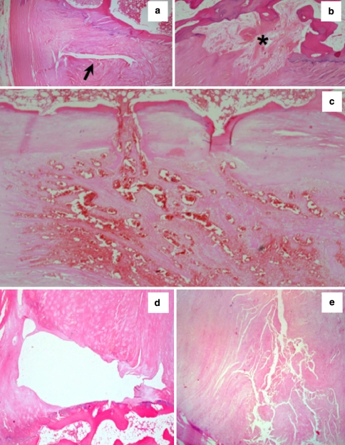
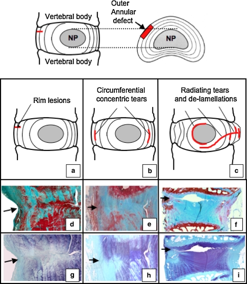
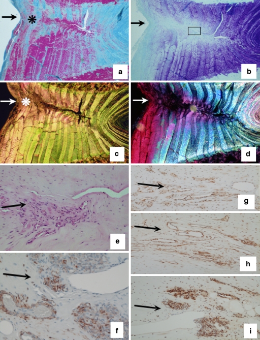
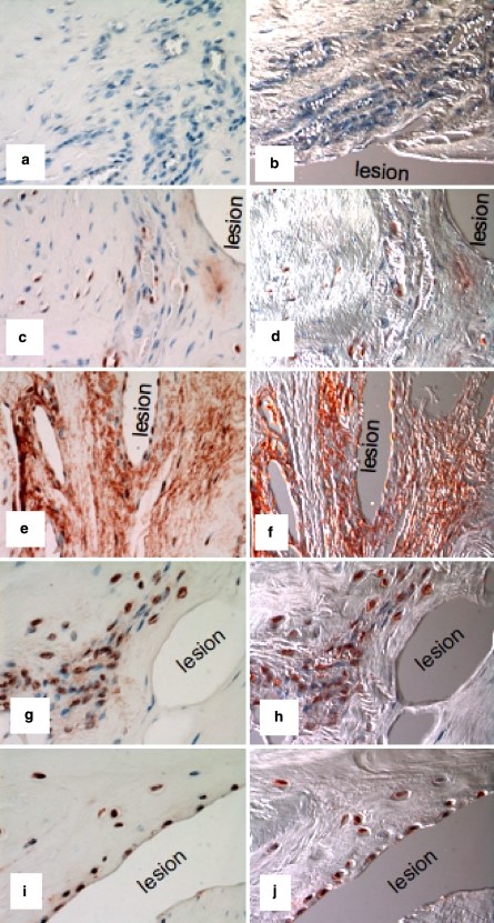
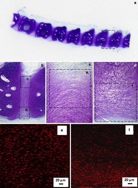
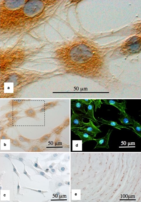
The objective of this study was to assess the impact of a landmark annular lesion model on our understanding of the etiopathogenesis of IVD degeneration and to appraise current IVD repairative strategies. A number of studies have utilised the Osti sheep model since its development in 1990. The experimental questions posed at that time are covered in this review, as are significant recent advances in annular repair strategies. The ovine model has provided important spatial and temporal insights into the longitudinal development of annular lesions and how they impact on other discal and paradiscal components such as the NP, cartilaginous end plates, zygapophyseal joints and vertebral bone and blood vessels. Important recent advances have been made in biomatrix design for IVD repair and in the oriented and dynamic culture of annular fibrochondrocytes into planar, spatially relevant, annular type structures. The development of hyaluronan hydrogels capable of rapid in situ gelation offer the possibility of supplementation of matrices with cells and other biomimetics and represent a significant advance in biopolymer design. New generation biological glues and self-curing acrylic formulations which may be augmented with slow delivery biomimetics in microcarriers may also find application in the non-surgical repair of annular defects. Despite major advances, significant technical challenges still have to be overcome before the biological repair of this intractable connective tissue becomes a realistic alternative to conventional surgical intervention for the treatment of chronic degenerate IVDs.


Figures

Fig. 1
Fig. 1
Morphology of rim lesions in H&E stained human IVDs. a Typical cleft type rim lesion (arrow), b repaired rim lesion (asterisk), c vasoproliferative rim lesion, d cystic rim lesion and e fragmented rim lesion
Fig. 2
Fig. 2
Schematic representation of the controlled rim lesions (top, arrow) used in the ovine model of experimental intervertebral disc degeneration and the defects which have been observed using this model (ac). Histological evaluation of vertical disc sections stained with Masson-Trichrome (df) to assess collagenous reorganisation and toluidine blue–fast green to delineate the focal loss of anionic proteoglycan in the vicinity of the initial lesion site (gi). Remodelling of the outer lesion consistent with a spontaneous repair response is evident in the outer lesion, however, experimental rim lesions are capable of propagating to form de-lamellations after 3 months (d, g) which may form circumferential concentric tears after 6 months (e, h) and radiating tears spanning from the original defect site to the contralateral annulus after 12 months (f, i)
Fig. 3
Fig. 3
Experimental ovine annular lesions demonstrating a spontaneous repair response in the outer annulus fibrosus (asterisk) after 3 months and lack of repair of the defect internally (ad). The non-healed defect is visible towards the right hand side of each photosegment, an arrow at the left hand side marks where the defect was initially made. a. Focal collagenous remodelling of outer AF spanning the original defect site (Masson Trichrome); b Focal loss of anionic proteoglycan along the plane of the experimental annular defect, (toluidine blue/fast green stain). c Re-organisation of collagen in the outer AF in response to the experimental annular defect (asterisk), (picrosirius red stain viewed under polarised light). d Polarised light view of segment (b) depicting similar detail to segment (c). e Cellular infiltration into a defect site 6 months post-operatively, (H&E). (f) Cells in the vicinity of a defect site stain positively for α-smooth muscle actin 6 months post-operatively. A large proportion of the cells which migrate into inner annular repair sites express FGF-2 (gi) and are associated with small blood vessels in regions of the AF undergoing remodelling consistent with an active repair response at least in the outer AF but not so in the inner AF; (g) 3 months; (h) 6 months; (i) 12 months post-operatively. The photosegments in ei are typical higher power views from an area approximating to the boxed region in segment b
Fig. 4
Fig. 4
Immunolocalisation of TGF-β in vertical sections of lesion affected ovine IVDs with a pan TGF-β Ab which detects all TGF-β isoforms. The areas shown are in the approximate area of the mid AF depicted by the boxed area in Fig. 2b. Negative control using equivalent concentration of mouse IgG in place of primary antibody (a, b); 3-month lesion (c, d); 6-month lesion (e, f); 12-month lesion (g, h); 26-month lesion (i, j). The photosegments depicted in the right hand column (b, d, f, h, j) are viewed with Nomarski DIC optics and are of the corresponding photosegments to their left (a, c, e, g, i), red blood cells are prominent in the DIC imaged blood vessels. The inner AF lesions depicted were incapable of undergoing repair despite the influx of cells and blood vessels to the lesion site and the demonstrated localisation of FGF-2 and TGF-β which are known to promote such processes
Fig. 5
Fig. 5
Perlecan has important roles to play in matrix assembly and the development of the annulus fibrosus and other discal components. Low and medium power longitudinal mid sagittal sections through a 12-week-old human foetal spine stained with toluidine blue–fast green to depict the cartilaginous rudiments and developing intervertebral discs (ad). The boxed area in (a) is presented at higher magnification in (b), as are the boxed areas in (b) in segments (c and d). Perlecan immunolocalisation in the boxed areas (e, f) is presented in segments (e, f), respectively by indirect fluorescent confocal microscopy using a rat anti perlecan primary mAb A7L6 to domain IV of perlecan and Alexa 594 conjugated donkey anti rat igG secondary Ab (red chromophore)
Fig. 6
Fig. 6
Perlecan provides cell-cell and cell-ECM interconnections important for cellular attachment and possibly mechanosensory processes. Immunolocalisation of pericellular perlecan in short-term primary monolayer culture of ovine AF cells using a primary mAb A76 to perlecan domain-I and horse radish conjuagated secondary antibodies and diaminobenzidene substrate for visualisation (brown chromogen), nuclei are counterstained with eosin (a, b). A negative control using equivalent concentration of mouse IgG in place of primary antibody is presented in (c). Localisation of the actin cytoskeleton layed down by the AF cells in monolayer culture using Phalloidin-oregon green, cell nuclei stained with DAPI (d). Immunolocalisation of perlecan in the pericellular matrix of AF cells in the native tissue (brown chromogen) using a mAb to perlecan domain IV (mAb A7L6) (e). Perlecan provides both cell–cell and cell–matrix connections which allow the monolayer cells to attach and spread out and also attaches the AF cells to the pericellular and interterritorial matrix in the native tissue

Similar articles

Cited by

References

    1. Abe T, Yamada H, Nakajima H, Kikuchi T, Takaishi H, Tadakuma T, et al. Repair of full-thickness cartilage defects using liposomal transforming growth factor-beta1. J Orthop Sci. 2003;8:92–101. doi: 10.1007/s007760300016. - DOI - PubMed
    1. Abreu JG, Ketpura NI, Reversade B, Robertis EM. Connective-tissue growth factor (CTGF) modulates cell signalling by BMP and TGF-beta. Nat Cell Biol. 2002;4:599–604. - PMC - PubMed
    1. Acosta FL, Jr, Lotz J, Ames CP. The potential role of mesenchymal stem cell therapy for intervertebral disc degeneration: a critical overview. Neurosurg Focus. 2005;19:E4. doi: 10.3171/foc.2005.19.3.5. - DOI - PubMed
    1. Agrawal A, Guttapalli A, Narayan S, Albert TJ, Shapiro IM, Risbud MV. Normoxic stabilization of HIF-1alpha drives glycolytic metabolism and regulates aggrecan gene expression in nucleus pulposus cells of the rat intervertebral disk. Am J Physiol Cell Physiol. 2007;293:C621–C631. doi: 10.1152/ajpcell.00538.2006. - DOI - PubMed
    1. Alini M, Li W, Markovic P, Aebi M, Spiro RC, Roughley PJ. The potential and limitations of a cell-seeded collagen/hyaluronan scaffold to engineer an intervertebral disc-like matrix. Spine. 2003;28:446–454. doi: 10.1097/00007632-200303010-00007. - DOI - PubMed

Publication types