Comparative Study
. 2008 Aug 2;7(8):1340-51.
doi: 10.1016/j.dnarep.2008.04.018. Epub 2008 Jun 26.

A comparison of BRCT domains involved in nonhomologous end-joining: introducing the solution structure of the BRCT domain of polymerase lambda

Affiliations
Comparative Study

A comparison of BRCT domains involved in nonhomologous end-joining: introducing the solution structure of the BRCT domain of polymerase lambda

Geoffrey A Mueller et al. DNA Repair (Amst). .

Abstract

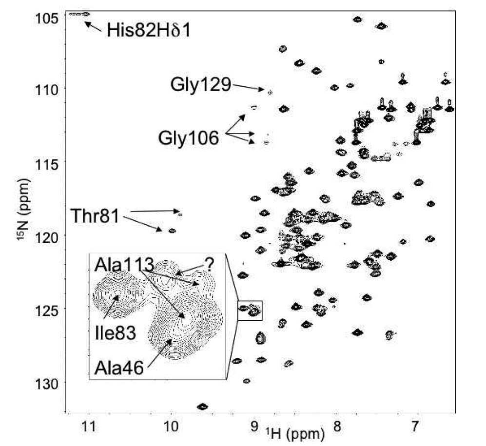
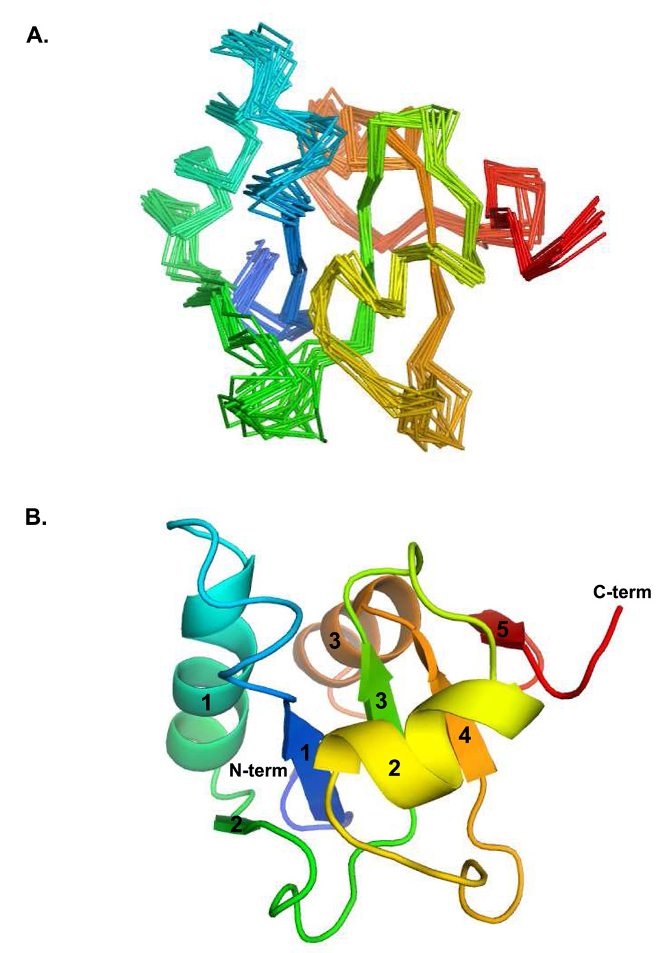
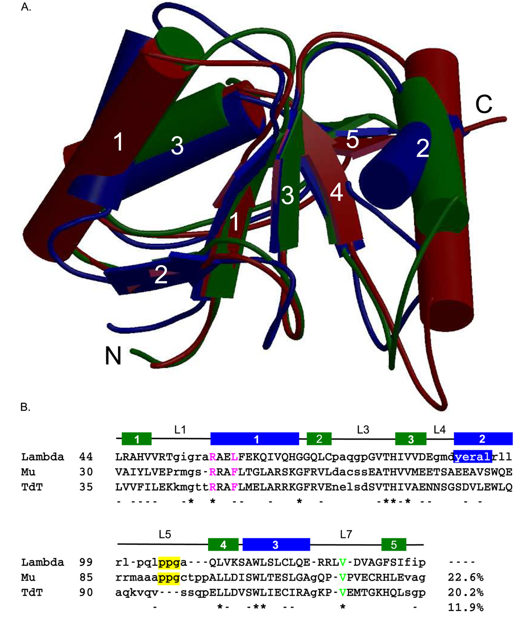
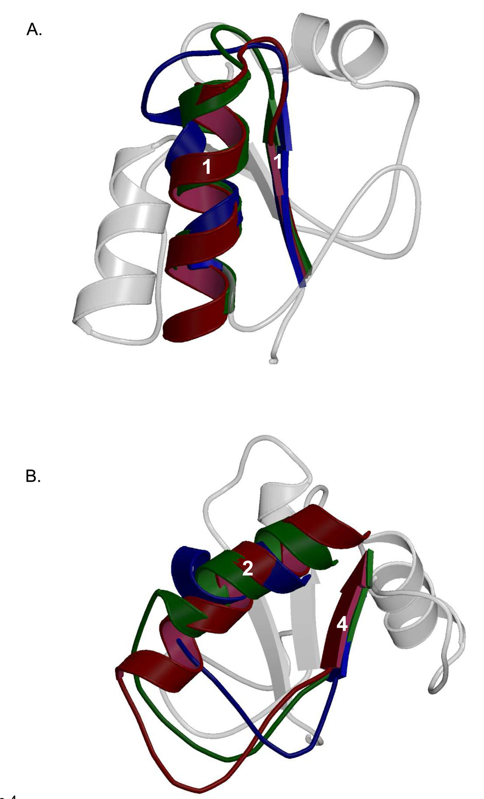
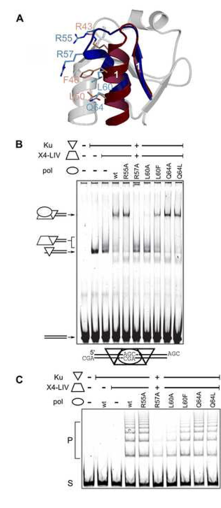
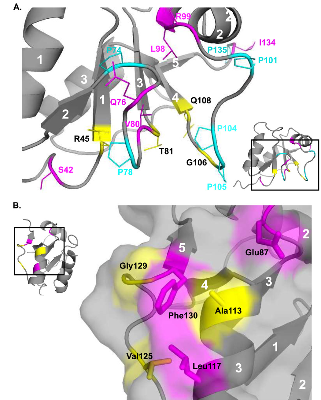
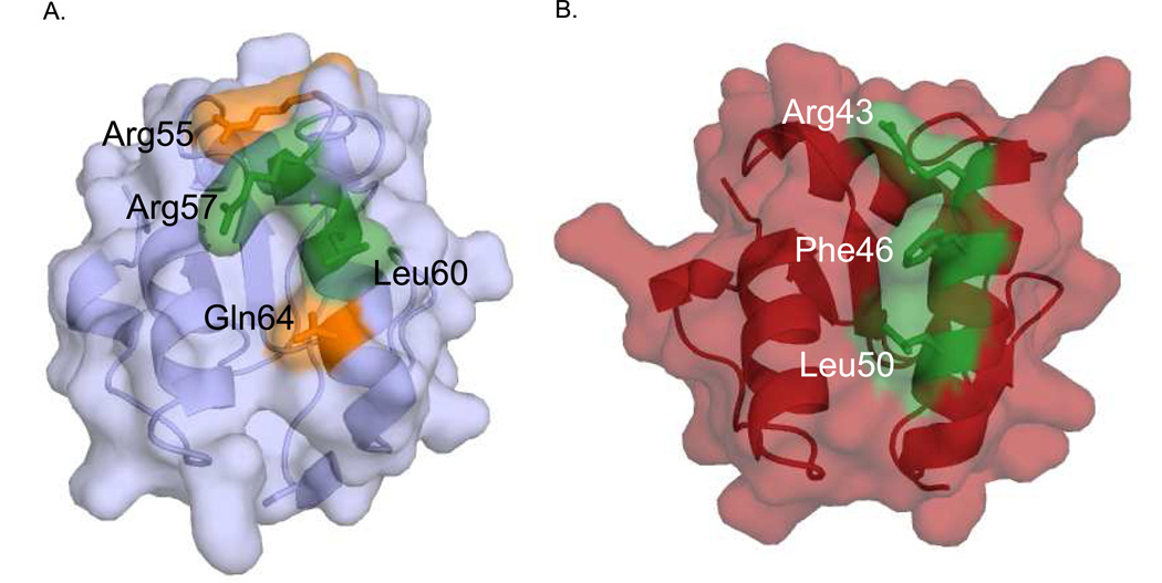
Three of the four family X polymerases, DNA polymerase lambda, DNA polymerase mu, and TdT, have been associated with repair of double-strand DNA breaks by nonhomologous end-joining. Their involvement in this DNA repair process requires an N-terminal BRCT domain that mediates interaction with other protein factors required for recognition and binding of broken DNA ends. Here we present the NMR solution structure of the BRCT domain of DNA polymerase lambda, completing the structural portrait for this family of enzymes. Analysis of the overall fold of the polymerase lambda BRCT domain reveals structural similarity to the BRCT domains of polymerase mu and TdT, yet highlights some key sequence and structural differences that may account for important differences in the biological activities of these enzymes and their roles in nonhomologous end-joining. Mutagenesis studies indicate that the conserved Arg57 residue of Pol lambda plays a more critical role for binding to the XRCC4-Ligase IV complex than its structural homolog in Pol mu, Arg43. In contrast, the hydrophobic Leu60 residue of Pol lambda contributes less significantly to binding than the structurally homologous Phe46 residue of Pol mu. A third leucine residue involved in the binding and activity of Pol mu, is nonconservatively replaced by a glutamine in Pol lambda (Gln64) and, based on binding and activity data, is apparently unimportant for Pol lambda interactions with the NHEJ complex. In conclusion, both the structure of the Pol lambda BRCT domain and its mode of interaction with the other components of the NHEJ complex significantly differ from the two previously studied homologs, Pol mu and TdT.


Figures

Figure 1
Figure 1
Annotated HSQC of [1H-15N]-Pol λ BRCT domain. Inset: Expanded view of degenerate peaks surrounding Ala113.
Figure 2
Figure 2
Ensemble and secondary structure of Pol λ BRCT domain. (A) The ensemble of structures shows good convergence with the exception of a few loops. (B) The core parallel β-sheet characteristic of BRCT domains is well determined. The lowest energy structure from the ensemble was used to generate this image, using PyMOL [49]. The secondary structural elements are shaded from blue to red; α-helices and β-strands are labeled numerically.
Figure 3
Figure 3
Structural alignment of the BRCT domains of the Family X polymerases. (A). Superposition of the BRCT domains from human Pol µ (PDB code 2DUN, maroon), TdT (PDB code 2COE, green), and Pol λ (PDB code 2JW5, blue). α-helices (cylinders) and β-strands (directional arrows) are labeled numerically. This figure was created using MolScript [50] and Raster3D [51] (B). Structure-based primary sequence alignment. The positions of the conserved secondary structural elements of the Pol λ BRCT domain are shown as follows: β-strands are drawn as green boxes and α-helices are drawn as blue boxes. Structurally homologous residues are in capital letters, while nonhomologous regions are shown in lower-case letters. Absolutely conserved residues are indicated by asterisks below the alignment, and nearly conserved residues are dashed. The PPG motif is boxed in yellow. The conserved valine residue required for protein stability is shown in green. Residues mutated for biochemical analysis of binding to NHEJ protein partners are in magenta.
Figure 4
Figure 4
Comparison of the structural differences seen in Family X BRCT domain loop regions. Secondary structural elements of Pol λ BRCT are shown in the background in light gray. (A). Superposition of the loop between β-strand 1 and α-helix 1 in Pol µ (PDB code 2DUN, maroon), TdT (PDB code 2COE, green), and Pol λ (PDB code 2JW5, blue). (B). Superposition of the loop between α-helix 2 and β-strand 4 in Pol µ (PDB code 2DUN, maroon), TdT (PDB code 2COE, green), and Pol λ (PDB code 2JW5, blue).
Figure 5
Figure 5
NHEJ activity of Pol λ BRCT domain mutants. (A). Pol λ (PDB code 2JW5), blue) superimposed with Pol µ (PDB code 2DUN, maroon). Residues in α-helix 1 crucial for interaction with core NHEJ factors in Pol µ (R43, F46, and L50, light red) are shown with sticks, relative to tested positions in Pol λ (R55, R57, L60 and Q64, light blue). Secondary structural elements of Pol λ BRCT are shown in the background in light gray. (B). EMSA analysis was performed in the presence of a 60 bp DNA duplex, Ku and the XRCC4-ligase IV (X4-LIV) complex and various full-length Pol λ proteins. . The composition of each species of distinct mobility is noted with cartoons at the left of the panel. C) Joining of a 300 bp substrate with three nucleotide AGC 3’ overhangs was performed in the presence of Ku, XRCC4-Ligase IV, and various full-length Pol λ constructs as noted. S, substrate, P, joined concatemer products.
Figure 6
Figure 6
Two views of slow motions in Pol λ BRCT domain. Residues that demonstrated Rex from CPMG experiments are colored magenta, and residues with multiple peaks in the 15N-1H HSQC are colored yellow. Proline residues are colored cyan. (A). Numerous residues in the vicinity of prolines that display either Rex or slow exchange cross-peaks. (B). A patch of surface-exposed residues that are mostly hydrophobic, and also show either Rex or slow exchange cross-peaks.
Figure 7
Figure 7
Molecular surface of Pol λ and Pol µ BRCT domains, highlighting important residues for NHEJ interactions. Pol λ (A) and Pol µ (B) are rendered, highlighting residues tested for NHEJ activity. Residues with activity similar to wildtype are colored orange and residues showing decreased binding and NHEJ activity are colored green.

References

    1. Braithwaite EK, Prasad R, Shock DD, Hou EW, Beard WA, Wilson SH. DNA polymerase lambda mediates a back-up base excision repair activity in extracts of mouse embryonic fibroblasts. J Biol Chem. 2005;280:18469–18475. - PubMed
    1. Srivastava DK, Berg BJ, Prasad R, Molina JT, Beard WA, Tomkinson AE, Wilson SH. Mammalian abasic site base excision repair. Identification of the reaction sequence and rate-determining steps. J Biol Chem. 1998;273:21203–21209. - PubMed
    1. Mahajan KN, Nick McElhinny SA, Mitchell BS, Ramsden DA. Association of DNA polymerase mu (pol mu) with Ku and ligase IV: role for pol mu in end-joining double-strand break repair. Mol Cell Biol. 2002;22:5194–5202. - PMC - PubMed
    1. Fan W, Wu X. DNA polymerase lambda can elongate on DNA substrates mimicking non-homologous end joining and interact with XRCC4-ligase IV complex. Biochem Biophys Res Commun. 2004;323:1328–1333. - PubMed
    1. Lee JW, Blanco L, Zhou T, Garcia-Diaz M, Bebenek K, Kunkel TA, Wang Z, Povirk LF. Implication of DNA polymerase lambda in alignment-based gap filling for nonhomologous DNA end joining in human nuclear extracts. J Biol Chem. 2004;279:805–811. - PubMed

Publication types