. 2008 Sep;39(1):83-94.
doi: 10.1016/j.mcn.2008.05.019. Epub 2008 Jun 7.

Synaptic adhesion-like molecules (SALMs) promote neurite outgrowth

Affiliations

Synaptic adhesion-like molecules (SALMs) promote neurite outgrowth

Philip Y Wang et al. Mol Cell Neurosci. 2008 Sep.

Abstract

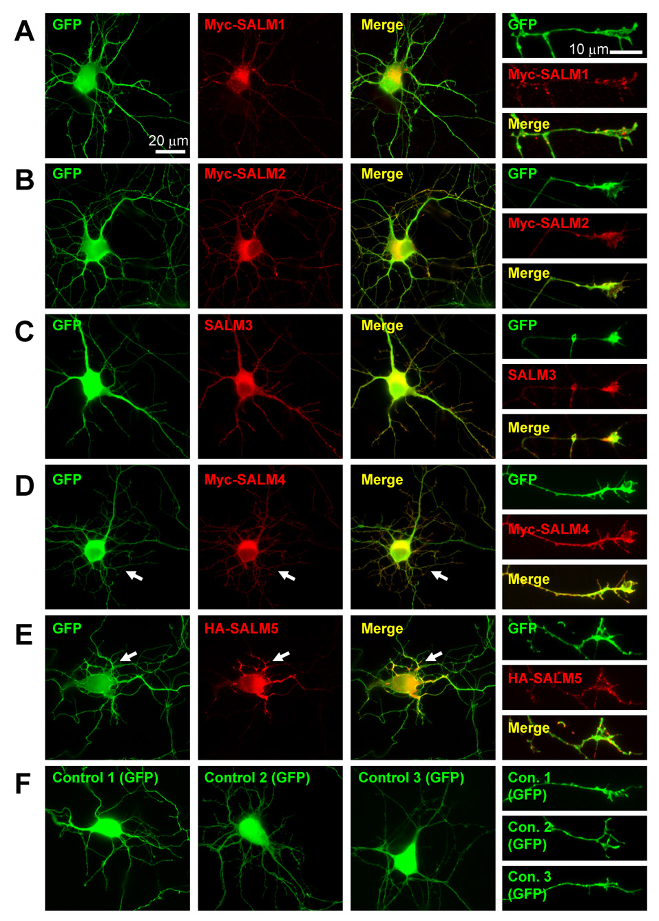
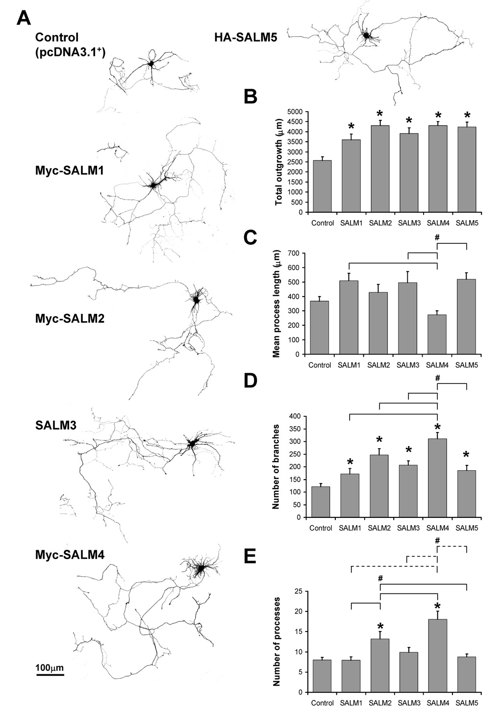
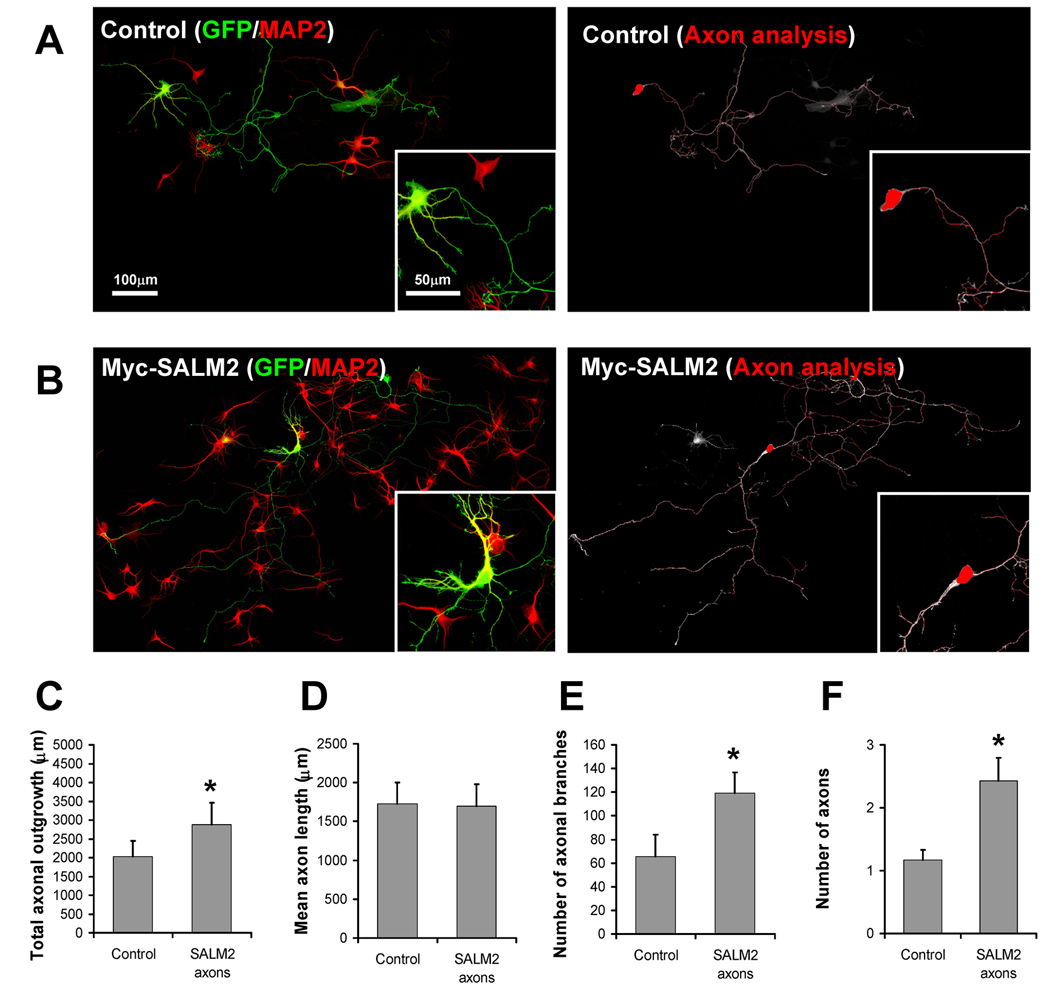
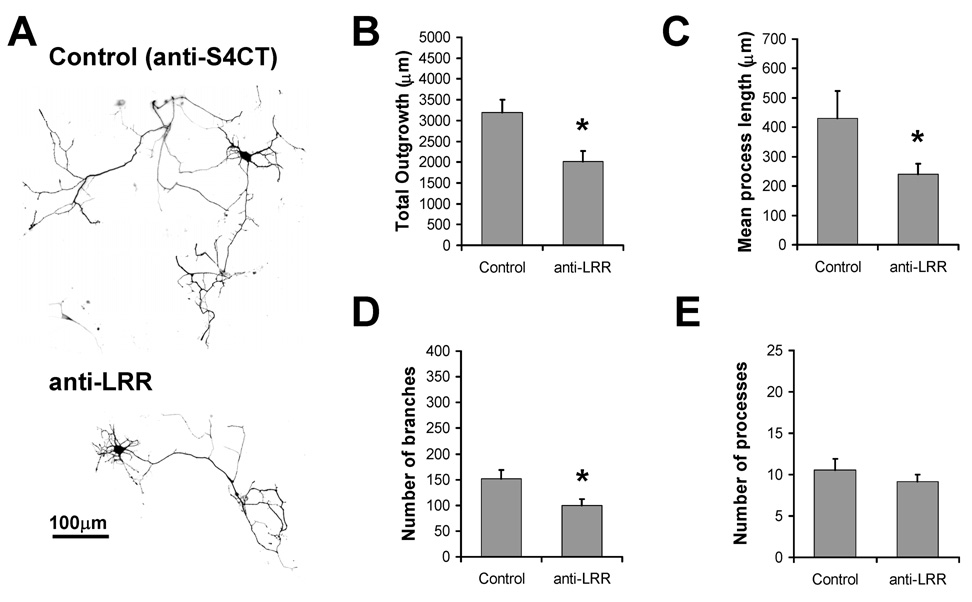
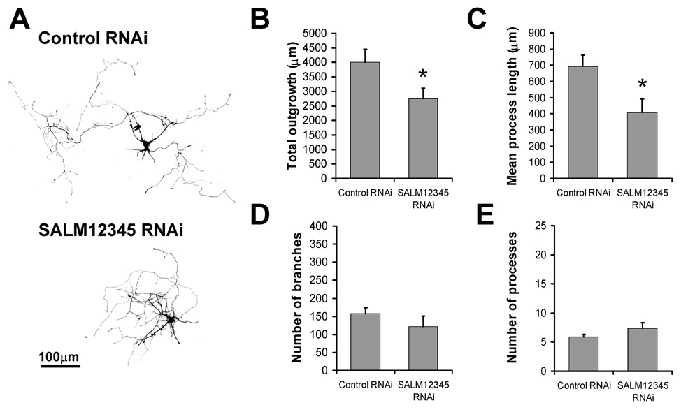
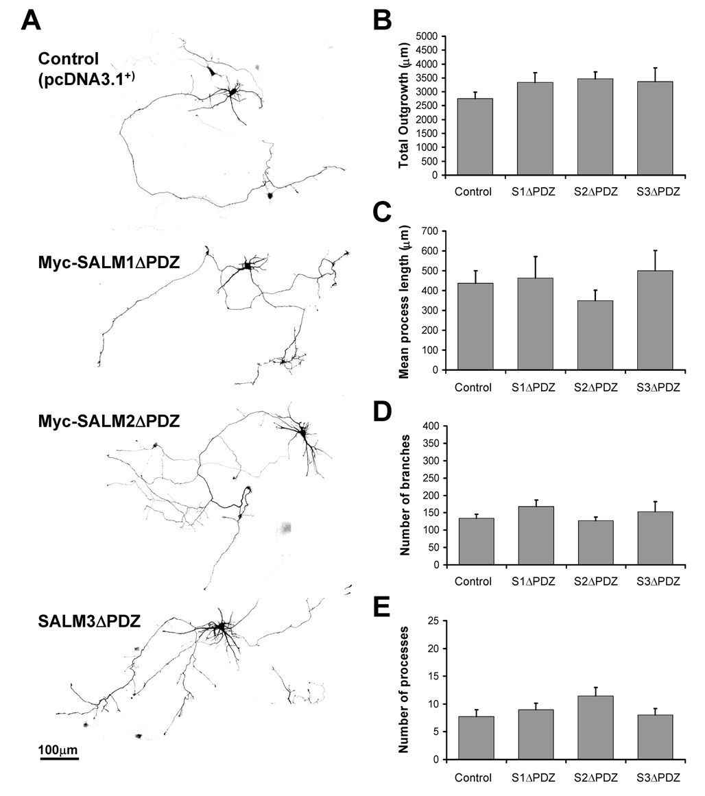
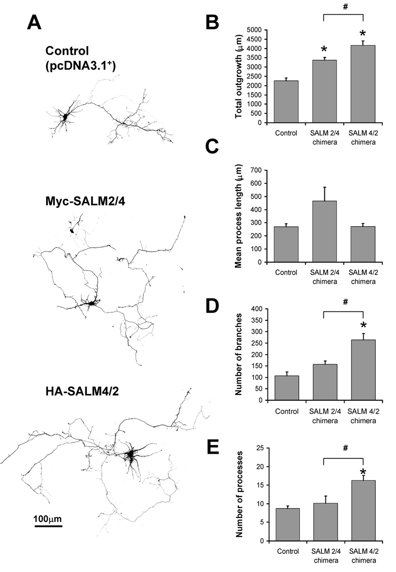
SALMs are a family of five adhesion molecules whose expression is largely restricted to the CNS. Initial reports showed that SALM1 functions in neurite outgrowth while SALM2 is involved in synapse formation. To investigate the function of SALMs in detail, we asked if all five are involved in neurite outgrowth. Expression of epitope-tagged proteins in cultured hippocampal neurons showed that SALMs are distributed throughout neurons, including axons, dendrites, and growth cones. Over-expression of each SALM resulted in enhanced neurite outgrowth, but with different phenotypes. Neurite outgrowth could be reduced by applying antibodies targeting the extracellular leucine rich regions of SALMs and with RNAi. Through over-expression of deletion constructs, we found that the C-terminal PDZ binding domains of SALMs 1-3 are required for most aspects of neurite outgrowth. In addition, by using a chimera of SALMs 2 and 4, we found that the N-terminus is also involved in neurite outgrowth.


Figures

Fig. 1
Fig. 1. Localization and morphological characteristics of transfected SALM proteins
DIV4 primary hippocampal cultures were co-transfected with SALM and GFP cDNAs. Neurons co-transfected with pcDNA3.1+ (empty vector) and GFP were used for control. Immunostaining was performed 48 hours later and the SALM localization and cell morphology were examined. Transfected SALMs localize throughout the cell body, axons, and dendrites. Representative examples of neurons transfected with SALMs 1–5 are shown in A–E, respectively. Scale bars, 20 µm. Transfected SALMs are enriched at growth cones (A–E, fourth column). Scale bars, 10 µm. Transfected SALM4 and SALM5, which do not contain PDZ-BDs, often exhibit distinct phenotypes as compared to SALMs 1–3. D, Myc-SALM4-transfected cells show a large increase in the number of short primary processes (arrows). HA-SALM5-transfected cells often display an increase in the number of primary processes crossing/overlapping with each other. E, Transfected SALM5 accumulates at these crossing points (arrows). Representative examples of GFP and pcDNA3.1+ co-transfected (control) cell body and growth cone morphology are shown in F.
Fig. 2
Fig. 2. SALMs promote neurite outgrowth
DIV4 primary hippocampal neurons were co-transfected with GFP and myc-SALM1, myc-SALM2, SALM3, myc-SALM4, HA-SALM5, or pcDNA 3.1+ vector (control). Immunostaining was performed 48 hours later, and neurite outgrowth was analyzed using Metamorph Neurite Outgrowth software (v7.0r3). Analysis was based on the transfected GFP signal. A, Representative examples of transfected neurons. B, Quantifications of neurite outgrowth: all five SALMs promote increases in total outgrowth, as compared to control, but do not promote increases in mean process length (C). The mean process length of myc-SALM4-transfected neurons is less than that of neurons transfected with myc-SALM1, SALM3, and HA-SALM5. D, All five SALMs promote increases in the number of branches, as compared to control. Additionally, myc-SALM4 promotes increases number of branches as compared to the other SALMs. E, Myc-SALM2 promotes increases in the number of processes, as compared to control, myc-SALM1, and HA-SALM5. Myc-SALM4 promotes increases in processes as compared to myc-SALM1, myc-SALM2, SALM3, HA-SALM5 (dotted lines) and control (n=15–17, values shown are mean +/− SEM, and analyzed by one-way ANOVA, * represents significance with respect to control, # represents significance with respect to other conditions, p<0.05). Scale bar, 100 µm.
Fig. 3
Fig. 3. SALM2 promotes axonal outgrowth
DIV4 primary hippocampal neurons were co-transfected with GFP and myc-SALM2 or pcDNA 3.1+ empty vector (control). Representative examples of control and myc-SALM2 transfected cells used for analysis are shown in A and B, respectively. Axons and dendrites were identified by morphological criteria and the dendritic marker MAP2 (A and B, left). Dendrites were digitally separated from the cell body, and the resulting axon-only images were analyzed for neurite outgrowth (A and B, right). Scale bars for composite images and insets 100 µm and 50 µm, respectively. Myc-SALM2 promotes an increase in total axonal outgrowth (C), number of axonal branches (E), and number of axons (F), as compared to controls. D, Myc-SALM2 does not promote increases in mean axon length (n=7, values shown are mean +/− SEM, and analyzed by unpaired students t-test, *p<0.05).
Fig. 4
Fig. 4. Application of antibodies directed to the extracellular LRR region of SALMs inhibits neurite outgrowth
DIV4 primary hippocampal neurons were transfected with GFP and treated by bath application with antibodies directed to the LRR region of SALM2 (anti-LRR) or to the C-terminus of SALM4 (anti-S4CT), as control. Immunostaining was performed 48 hours later, and neurite outgrowth was analyzed using Metamorph. A, Representative examples of transfected/treated neurons. Neurons treated with anti-LRR showed a significant decrease in total outgrowth (B), mean process length (C) and number of branches (D), as compared to the control condition. Anti-LRR treatment did not have an effect on the number of processes (E). (n=15, values shown are mean +/− SEM, and analyzed by unpaired students t-test, *p<0.05. Scale bar, 100 µm.)
Fig. 5
Fig. 5. RNAi knock-down of SALM expression reduces neurite outgrowth
Individual RNAi plasmid constructs were designed and generated for SALMs 1–5 (Invitrogen custom services). To knockdown the expression of endogenous SALM proteins, DIV2 neurons were quintuple transfected with SALMs 1–5 RNAi constructs (SALM12345 RNAi), and analyzed four days later (DIV6). A, Representative examples of transfected cells. GFP-positive RNAi-transfected cells showed a significant decrease in total outgrowth and mean process length (B and C, respectively). The number of branches and processes were not significantly different, compared to control (D and E, respectively). (n=10, values shown are mean +/− SEM, and analyzed by unpaired students t-test, *p<0.05. Scale bar, 100 µm.)
Fig. 6
Fig. 6. Neurite outgrowth is mediated by the PDZ domains of SALMs
Primary hippocampal neurons (DIV4) were co-transfected with GFP and myc-SALM1ΔPDZ, myc-SALM2ΔPDZ, SALM3ΔPDZ, or pcDNA 3.1+ vector (control). Immunostaining was performed 48 hours later, and neurite outgrowth was analyzed using Metamorph. A, Representative examples of transfected neurons. Transfection of myc-SALM1ΔPDZ, myc-SALM2ΔPDZ, or SALM3ΔPDZ do not promote increases in total outgrowth (B), mean process length (C), number of branches (D), or the number of processes (E). (n=10, values shown are mean +/− SEM, and analyzed by one-way ANOVA, *p<0.05. Scale bar, 100 µm.)
Fig. 7
Fig. 7. Neurite outgrowth phenotype is determined by both the N-and C-termini of SALMs
We generated SALM2/SALM4 chimera constructs in which the N-termini and C-termini of SALM2 and SALM4 were switched. Myc-SALM2/4 contains the N-terminus of SALM2 and the C-terminus of SALM4, while HA-SALM4/2 contains the N-terminus of SALM4 and the C-terminus of SALM2. Chimera constructs were individually co-transfected with GFP at DIV4 and analyzed 48 hours later. A, Representative examples of pcDNA3.1+ (control) and chimera-transfected cells. Both Myc-SALM2/4 and HA-SALM4/2 exhibited increases in total outgrowth (B), but not mean process length (C), as compared to control. HA-SALM4/2 promoted increases in number of branches (D) and process number (E), as compared to both control and Myc-SALM2/4. The HA-SALM4/2-mediated increase in process number is similar in phenotype to the increase seen with SALM4, indicating that the N-terminus of SALM4 confers this property (n=10, values shown are mean +/− SEM, and analyzed by one-way ANOVA, * represents significance with respect to control, # represents significance with respect to other conditions, p<0.05. Scale bar, 100 µm).

Similar articles

Cited by

References

    1. Bixby JL, Pratt RS, Lilien J, Reichardt LF. Neurite outgrowth on muscle cell surfaces involves extracellular matrix receptors as well as Ca2+-dependent and -independent cell adhesion molecules. Proc Natl Acad Sci U S A. 1987;84:2555–2559. - PMC - PubMed
    1. Bixby JL, Zhang R. Purified N-cadherin is a potent substrate for the rapid induction of neurite outgrowth. J Cell Biol. 1990;110:1253–1260. - PMC - PubMed
    1. Charych EI, Akum BF, Goldberg JS, Jornsten RJ, Rongo C, Zheng JQ, Firestein BL. Activity-independent regulation of dendrite patterning by postsynaptic density protein PSD-95. J Neurosci. 2006;26:10164–10176. - PMC - PubMed
    1. Chen Y, Aulia S, Li L, Tang BL. AMIGO and friends: an emerging family of brain-enriched, neuronal growth modulating, type I transmembrane proteins with leucine-rich repeats (LRR) and cell adhesion molecule motifs. Brain Res Rev. 2006;51:265–274. - PubMed
    1. Craig AM, Banker G. Neuronal polarity. Annu Rev Neurosci. 1994;17:267–310. - PubMed

Publication types

MeSH terms