. 2008 Aug;135(2):610-20.
doi: 10.1053/j.gastro.2008.05.032. Epub 2008 May 15.

Myosin light chain kinase is central to smooth muscle contraction and required for gastrointestinal motility in mice

Affiliations

Myosin light chain kinase is central to smooth muscle contraction and required for gastrointestinal motility in mice

Wei-Qi He et al. Gastroenterology. 2008 Aug.

Abstract

Background & aims: Smooth muscle is essential for maintaining homeostasis for many body functions and provides adaptive responses to stresses imposed by pathologic disorders. Identified cell signaling networks have defined many potential mechanisms for initiating smooth muscle contraction with or without myosin regulatory light chain (RLC) phosphorylation by myosin light chain kinase (MLCK). We generated tamoxifen-inducible and smooth muscle-specific MLCK knockout (KO) mice and provide direct loss-of-function evidence that shows the primary importance of MLCK in phasic smooth muscle contractions.

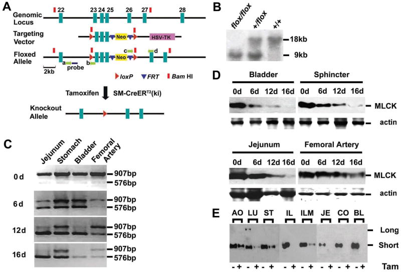
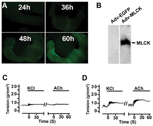
Methods: We used the Cre-loxP system to establish Mlck floxed mice in which exons 23, 24, and 25 were flanked by 2 loxP sites. Smooth muscle-specific MLCK KO mice were generated by crossing Mlck floxed mice with SM-CreER(T2) (ki) mice followed by tamoxifen treatment. The phenotype was assessed by histologic, biochemical, molecular, cell biological, and physiologic analyses.

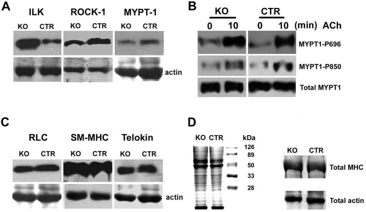
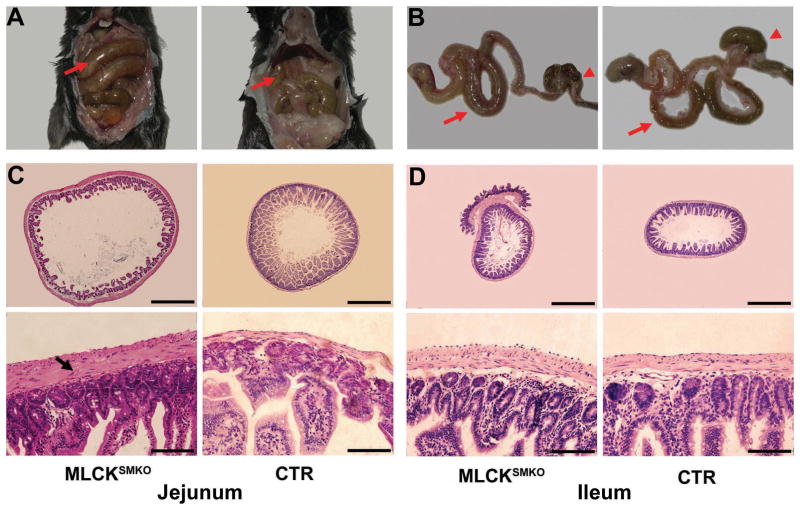
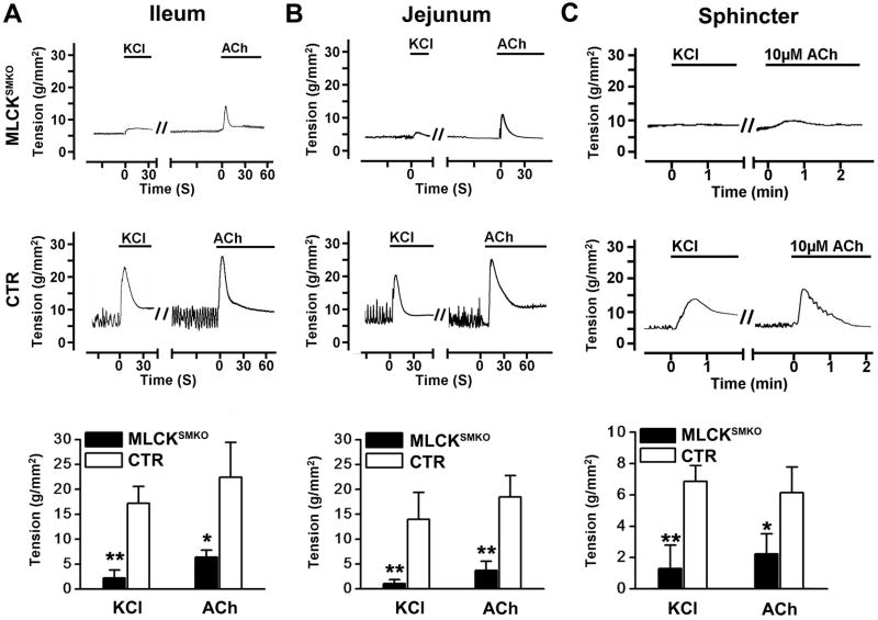
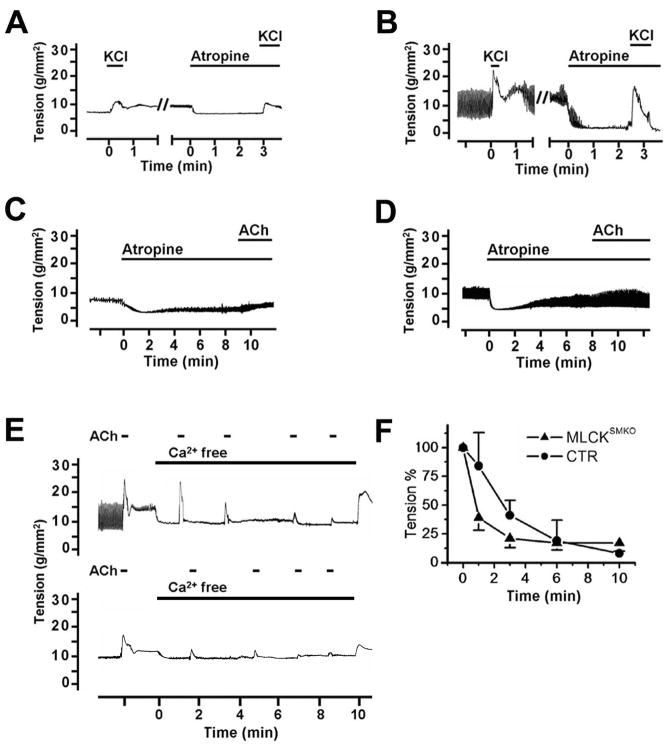
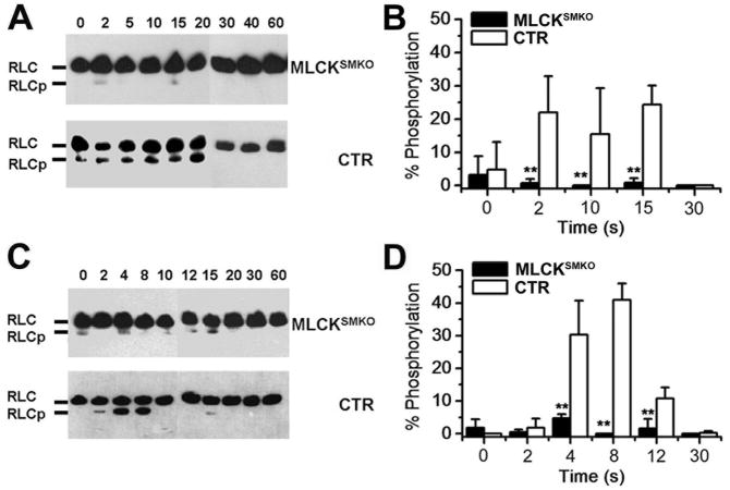
Results: Targeted deletion of MLCK in adult mouse smooth muscle resulted in severe gut dysmotility characterized by weak peristalsis, dilation of the digestive tract, and reduction of feces excretion and food intake. There was also abnormal urinary bladder function and lower blood pressure. Isolated muscles showed a loss of RLC phosphorylation and force development induced by K(+)-depolarization. The kinase knockout also markedly reduced RLC phosphorylation and force development with acetylcholine which activates Ca(2+)-sensitizing signaling pathways.

Conclusions: MLCK and its phosphorylation of RLC are required physiologically for smooth muscle contraction and are essential for normal gastrointestinal motility.


Conflict of interest statement

Conflicts of interest: There are no conflicts of interest to disclose.

Figures

Figure 1
Figure 1
Targeted disruption of Mlck gene in smooth muscle. (A) Schematic representation of Mlck smooth muscle-specific knockout strategy. The 8.9-kb genomic DNA fragment containing Mlck exons 23–25 was subcloned from 129/sv BAC using gap repair. The floxed Neo cassette was targeted upstream of exon 23 and excision of the floxed Neo cassette left behind a single loxP site (arrowheads in red) at the targeted locus. The single PGK-Neo cassette flanked by FRT sites (arrowheads in blue) and a downstream loxP site, was then introduced downstream of exon 25. The Neo cassette contains a BamHI site (blocks in red) which is favorable for Southern blot analysis. The floxed allele (Mlckflox) was formed after homologous recombination in ES cells. Mice containing the floxed allele were crossed with SM-CreERT2 (ki) mice that express a tamoxifen-activated Cre recombinase to generate Mlck+/flox; SM-CreERT2 and Mlckflox/flox; SM-CreERT2 mice. The ablation of exons 23–25 was induced by tamoxifen injection. The probe used for Southern blot analysis is shown as a solid blue bar, and the locations of the PCR primers a–d are indicated by green arrows. (B) Tail DNA isolated from homozygous (flox/flox) floxed, heterozygous (+/flox) and wild-type (+/+) mice was digested with BamHI and analyzed by Southern blot. The wild-type and floxed allele yield 18kb and 9kb fragments respectively. (C) RT-PCR assay for MLCK mRNA. Various smooth muscle tissues were collected from MLCKSMKO mice induced with tamoxifen at different time points. The mRNA containing exons 23–25 was amplified by RT-PCR. The products in size of 907bp and 576bp reflect wild-type and mutated MLCK respectively. (D) Western blots of MLCK in tamoxifen-treated tissues collected at indicated days (d). Total actin stained with Coomassie Brilliant Blue G-250 was used as protein loading control. Tissues included the urinary bladder, internal anal sphincter, jejunum and femoral artery. (E) Western blots of MLCK protein from floxed mice treated with tamoxifen (Tam+) or its vehicle (Tam-) for 16 days. The amount of loaded protein was normalized by total actin control. AO, aorta; LU, lung; ST, stomach; IL, ileum; ILM, ileum mucosa; JE, jejunum; CO, colon; BL, bladder.
Figure 2
Figure 2
Expression of contractile and related regulatory proteins in jejunum from MLCKSMKO mice. Western blot of ILK, ROCK1, MYPT1, RLC, SM-MHC and telokin (A and C) from MLCKSMKO (KO) and CTR jejunum. The samples were resolved by separate SDS-PAGE and total actin stained with Coomassie brilliant blue was used as loading control. MYPT1 phosphorylations at residues 696 and 850 in response to ACh were measured by anti-MYPT1-P696 and anti-MYPT1-P850 antibody (B). Ileal total proteins (30 μg) in MLCKSMKO and CTR were resolved by SDS-PAGE and stained with Coomassie brilliant blue (left side of D). To visualize more clearly the total myosin heavy chain and actin, we performed electrophoresis with different amounts of protein loading (100 μg) for comparisons of KO and CTR samples (right side of D).
Figure 3
Figure 3
Knockout of smooth muscle MLCK attenuates gastrointestinal motility. (A) Time course for food intake (left) and feces excretion (right) after tamoxifen treatment. Days were numbered with the start of tamoxifen injection. The value of each point represents the mean±SEM of MLCKSMKO and the CTR mice (n=3 each group). (B) Representative spontaneous contractions are shown for ileal segments from MLCKSMKO (left) and CTR (right) mice. (C) Quantification of the contraction amplitudes (left) and frequencies (right) are shown. (D) Tail blood pressure of MLCKSMKO and CTR mice was measured 15–16 days after beginning of tamoxifen injection (top). Micturition assay was performed at day 15. The total area of urine spots per hour (middle) and total number of urine spots per hour (bottom) were quantified. Bars represent means ± SEM, n=3–9, **p<0.01 (t-test).
Figure 4
Figure 4
Abnormal gastrointestinal tract of MLCKSMKO mice. Appearance of gastrointestinal tract of a MLCKSMKO and a CTR mouse in situ (A) and out of the body (B). Note the dilated jejunum (arrows) and compacted feces in caecum and colon (arrowheads) in the MLCKSMKO mouse. Histological examination of the small intestinal tract shows tissue changes with MLCK knockout in smooth muscle. Transverse sections of jejunum (C) and ileum (D) from MLCKSMKO and CTR mice were stained with hematoxylin and eosin. The lumen of the MLCKSMKO jejunum has a larger diameter with rare intestinal villus and a hypertrophic smooth muscle layer (indicated by an arrow). The morphology of ileum from MLCKSMKO mice appears normal except for the hypertrophic smooth muscle layer. Scale bars for C and D: upper row, 500 μm; lower row, 50 μm.
Figure 5
Figure 5
Contractions in intestinal smooth muscle and internal anal sphincter (IAS). Representative recordings of ileum (A) and jejunum (B), and IAS (C) from MLCKSMKO (upper row) and CTR (middle row) mice treated with 87 mM KCl or 1μM ACh. Bars show duration of stimulation. Quantification of contraction responses to KCl and ACh (bottom row) for ileum (A), and jejunum (B) and IAS (C). Bars represent means±SEM, n=4–10, **p<0.01, *p<0.05 (t-test).
Figure 6
Figure 6
Effects of atropine and Ca2+ depletion on smooth muscle contractility. KCl-induced contraction was not blocked by atropine (1μM) in ileal smooth muscle from MLCKSMKO (A) or CTR (B) mice. ACh (1μM) induced contractions in ileum from MLCKSMKO (C) and control (D) mice were inhibited by atropine. Depletion of Ca2+ in ileal strips by 1 mM EGTA inhibited the contractile responses to repeated exposure to ACh (E). Quantitation of contractile responses of MLCKSMKO and CTR muscles to calcium depletion were normalized (F). Values are means±SEM (n=3–4).
Figure 7
Figure 7
Expression of exogenous MLCK partially restores contraction of MLCKSMKO smooth muscle. (A) A jejunum smooth muscle strip (6 mm) was infected with MLCK-expressing adenovirus (Adv-MLCK) and cultured up to 60 h. The GFP expression driven by IRES as a tag of MLCK expression in a smooth muscle strip was examined by a dissecting fluorescence microscopy. (B) Exogenous MLCK was detected by Western blot in strips collected at 60 h. The two panels shown were from the same gel, but different lanes. Typical force recordings in response to 87 mM KCl or 1 μM ACh are shown for MLCK-deficient muscle infected with (C) Adv-GFP control virus or (D) Adv-MLCK virus.
Figure 8
Figure 8
Time course of RLC phosphorylation in ileal smooth muscle from MLCKSMKO and CTR mice. RLC phosphorylation was measured in quick-frozen ileum smooth muscles from MLCKSMKO and control mice treated with 87 mM KCl (A) or 1μM ACh (C) as shown by representative Western blots of glycerol/urea PAGE gels. B and D show quantitation of RLC phosphorylation with KCl and ACh treatments, respectively. Bars represent means±SEM, n=4–10, **p<0.01 (t-test).

Similar articles

Cited by

References

    1. Kamm KE, Stull JT. Dedicated myosin light chain kinases with diverse cellular functions. J Biol Chem. 2001;276:4527–4530. - PubMed
    1. Somlyo AP, Somlyo AV. Ca2+-sensitivity of smooth and non-muscle myosin II: modulation by G Proteins, kinases and myosin phosphatase. Physiological Rev. 2003;83:1325–1358. - PubMed
    1. Murthy KE. Signaling for contraction and relaxation in smooth muscle of the gut. Annu Rev Physiol. 2006;68:11.1–11.30. - PubMed
    1. Ito M, Nakano T, Erdödi F, et al. Myosin phosphatase: structure, regulation and function. Mol Cell Biochem. 2004;259:197–209. - PubMed
    1. Somlyo AP, Somlyo AV. Signal transduction and regulation in smooth muscle. Nature. 1994;372:231–236. - PubMed

Publication types

MeSH terms