

Role of oxidative stress in the induction of metallothionein-2A and heme oxygenase-1 gene expression by the antineoplastic agent gallium nitrate in human lymphoma cells
- PMID: 18586083
- PMCID: PMC2610863
- DOI: 10.1016/j.freeradbiomed.2008.05.031
Role of oxidative stress in the induction of metallothionein-2A and heme oxygenase-1 gene expression by the antineoplastic agent gallium nitrate in human lymphoma cells
Abstract
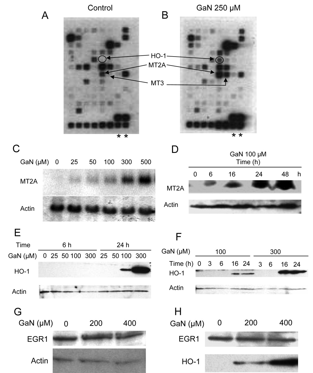
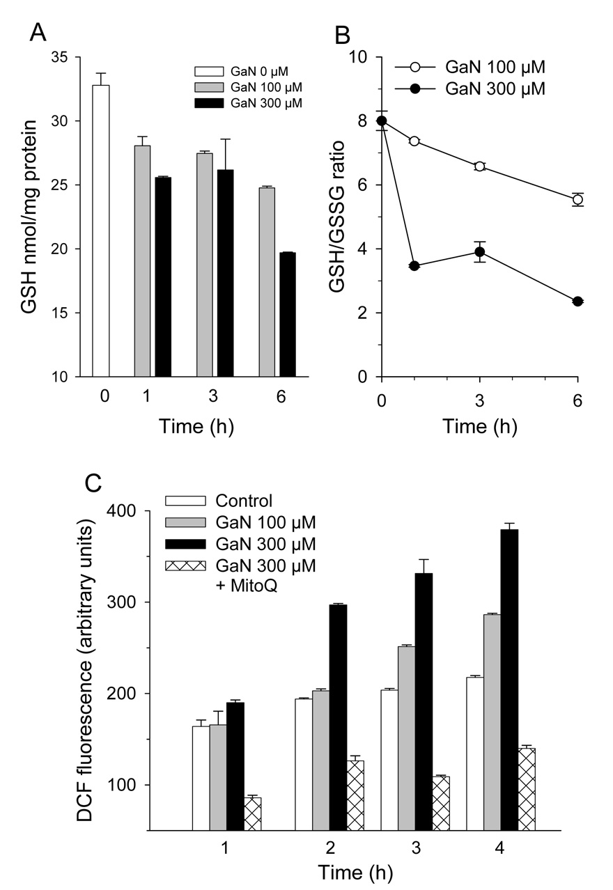
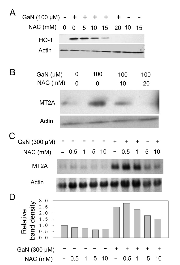
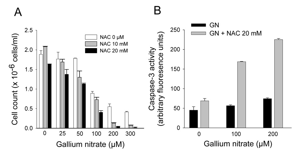
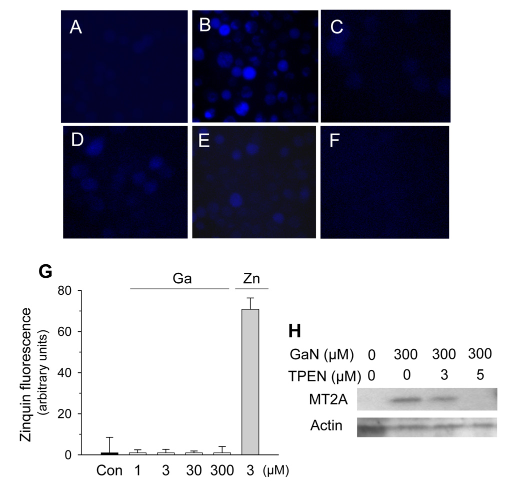
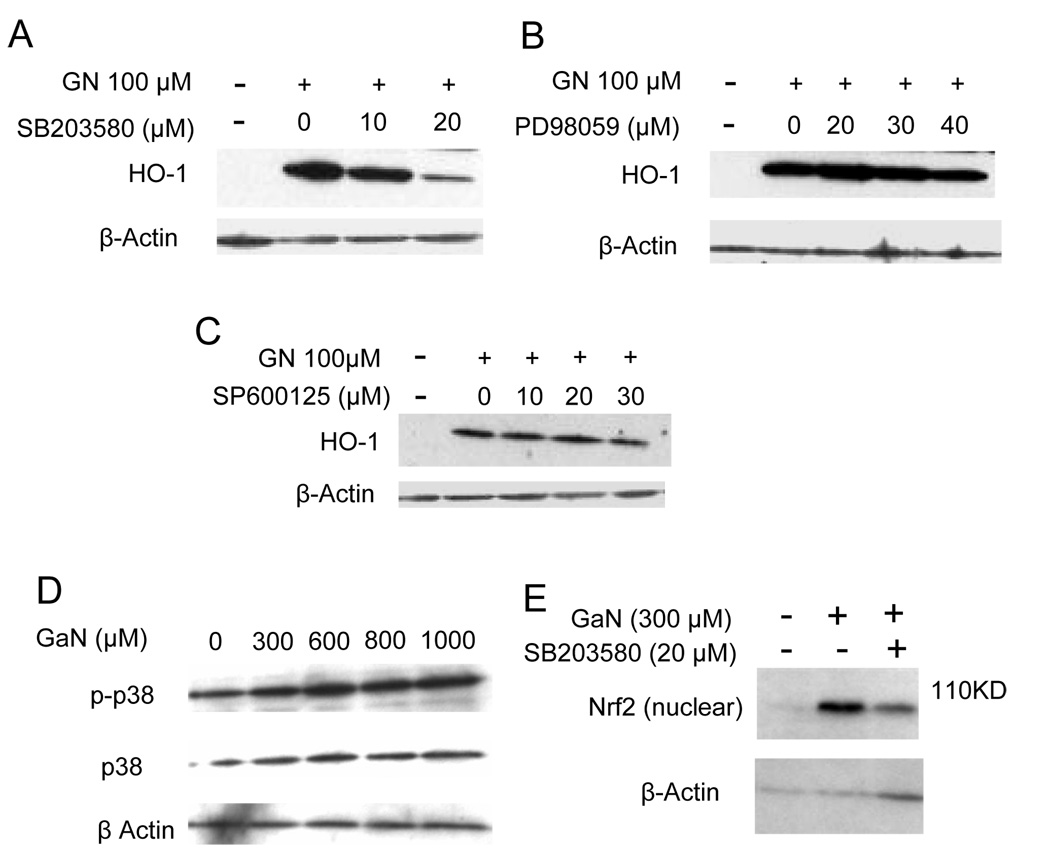
The mechanisms of action of gallium nitrate, an antineoplastic drug, are only partly understood. Using a DNA microarray to examine genes induced by gallium nitrate in CCRF-CEM cells, we found that gallium increased metallothionein-2A (MT2A) and heme oxygenase-1 (HO-1) gene expression and altered the levels of other stress-related genes. MT2A and HO-1 were increased after 6 and 16 h of incubation with gallium nitrate. An increase in oxidative stress, evidenced by a decrease in cellular GSH and GSH/GSSG ratio, and an increase in dichlorodihydrofluorescein (DCF) fluorescence, was seen after 1-4 h of incubation of cells with gallium nitrate. DCF fluorescence was blocked by the mitochondria-targeted antioxidant mitoquinone. N-Acetyl-L-cysteine blocked gallium-induced MT2A and HO-1 expression and increased gallium's cytotoxicity. Studies with a zinc-specific fluoroprobe suggested that gallium produced an expansion of an intracellular labile zinc pool, suggesting an action of gallium on zinc homeostasis. Gallium nitrate increased the phosphorylation of p38 mitogen-activated protein kinase and activated Nrf-2, a regulator of HO-1 gene transcription. Gallium-induced Nrf-2 activation and HO-1 expression were diminished by a p38 MAP kinase inhibitor. We conclude that gallium nitrate induces cellular oxidative stress as an early event which then triggers the expression of HO-1 and MT2A through different pathways.
Figures
Similar articles
-
Gene expression analysis of gallium-resistant and gallium-sensitive lymphoma cells reveals a role for metal-responsive transcription factor-1, metallothionein-2A, and zinc transporter-1 in modulating the antineoplastic activity of gallium nitrate.Mol Cancer Ther. 2007 Feb;6(2):633-43. doi: 10.1158/1535-7163.MCT-06-0557. Mol Cancer Ther. 2007. PMID: 17308060
-
Development of gallium compounds for treatment of lymphoma: gallium maltolate, a novel hydroxypyrone gallium compound, induces apoptosis and circumvents lymphoma cell resistance to gallium nitrate.J Pharmacol Exp Ther. 2007 Sep;322(3):1228-36. doi: 10.1124/jpet.107.126342. Epub 2007 Jun 28. J Pharmacol Exp Ther. 2007. PMID: 17600139
-
Diallyl sulfide induces heme oxygenase-1 through MAPK pathway.Arch Biochem Biophys. 2004 Dec 15;432(2):252-60. doi: 10.1016/j.abb.2004.09.024. Arch Biochem Biophys. 2004. PMID: 15542064
-
Gallium Complexes as Anticancer Drugs.Met Ions Life Sci. 2018 Feb 5;18:/books/9783110470734/9783110470734-016/9783110470734-016.xml. doi: 10.1515/9783110470734-016. Met Ions Life Sci. 2018. PMID: 29394029 Review.
-
Gallium compounds as antineoplastic agents.Curr Opin Oncol. 2004 Nov;16(6):547-52. doi: 10.1097/01.cco.0000142071.22226.d2. Curr Opin Oncol. 2004. PMID: 15627016 Review.
Cited by
-
ptk2 and mt2a Genes Expression in Gastritis and Gastric Cancer Patients with Helicobacter pylori Infection.Can J Gastroenterol Hepatol. 2022 Aug 25;2022:8699408. doi: 10.1155/2022/8699408. eCollection 2022. Can J Gastroenterol Hepatol. 2022. PMID: 36060520 Free PMC article.
-
Medical applications and toxicities of gallium compounds.Int J Environ Res Public Health. 2010 May;7(5):2337-61. doi: 10.3390/ijerph7052337. Epub 2010 May 10. Int J Environ Res Public Health. 2010. PMID: 20623028 Free PMC article. Review.
-
Methylmercury induces acute oxidative stress, altering Nrf2 protein level in primary microglial cells.Toxicol Sci. 2010 Aug;116(2):590-603. doi: 10.1093/toxsci/kfq126. Epub 2010 Apr 26. Toxicol Sci. 2010. PMID: 20421342 Free PMC article.
-
The cytotoxicity of gallium maltolate in glioblastoma cells is enhanced by metformin through combined action on mitochondrial complex 1.Oncotarget. 2020 Apr 28;11(17):1531-1544. doi: 10.18632/oncotarget.27567. eCollection 2020 Apr 28. Oncotarget. 2020. PMID: 32391122 Free PMC article.
-
Crystal structure and chemotherapeutic efficacy of the novel compound, gallium tetrachloride betaine, against breast cancer using nanotechnology.Tumour Biol. 2016 Aug;37(8):11025-38. doi: 10.1007/s13277-016-4969-2. Epub 2016 Feb 19. Tumour Biol. 2016. PMID: 26894603
References
-
- Collery P, Keppler B, Madoulet C, Desoize B. Gallium in cancer treatment. Crit Rev. Oncol. Hematol. 2002;42:283–296. - PubMed
-
- Chitambar CR. Gallium compounds as antineoplastic agents. Curr. Opin. Oncol. 2004;16:547–552. - PubMed
-
- Straus DJ. Gallium nitrate in the treatment of lymphoma. Semin. Oncol. 2003;30:25–33. - PubMed
-
- Einhorn L. Gallium nitrate in the treatment of bladder cancer. Semin. Oncol. 2003;30:34–41. - PubMed
-
- Keller J, Bartolucci A, Carpenter JT, Jr, Feagler J. Phase II evaluation of bolus gallium nitrate in lymphoproliferative disorders: A Southeastern Cancer Study Group trial. Cancer Treat. Rep. 1986;70:1221–1223. - PubMed
Publication types
MeSH terms
Substances
Grants and funding
LinkOut - more resources
Full Text Sources
Medical
