. 2008 Aug;45(2):250-60.
doi: 10.1016/j.yjmcc.2008.05.010. Epub 2008 May 27.

Darbepoetin alfa exerts a cardioprotective effect in autoimmune cardiomyopathy via reduction of ER stress and activation of the PI3K/Akt and STAT3 pathways

Affiliations

Darbepoetin alfa exerts a cardioprotective effect in autoimmune cardiomyopathy via reduction of ER stress and activation of the PI3K/Akt and STAT3 pathways

Weike Mao et al. J Mol Cell Cardiol. 2008 Aug.

Abstract

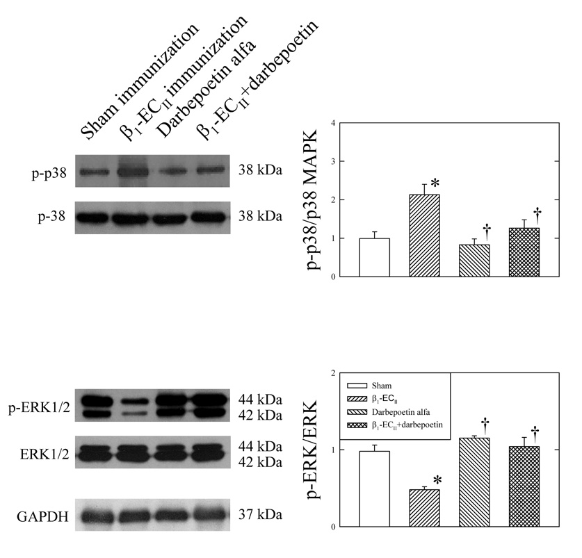
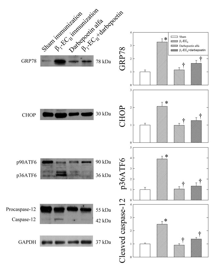
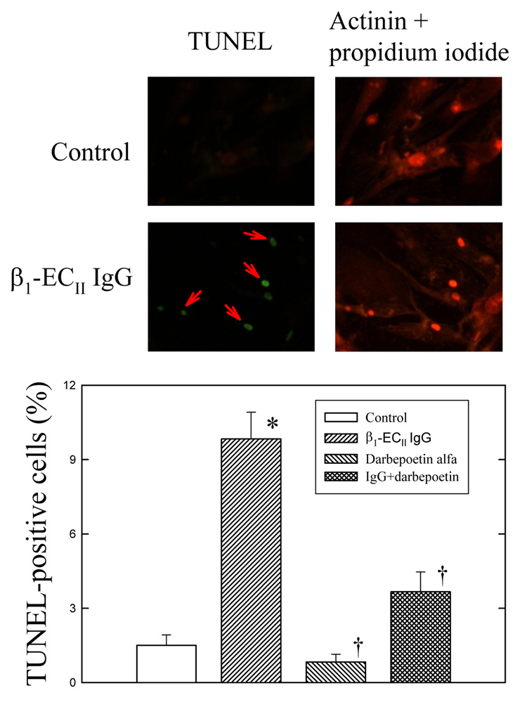
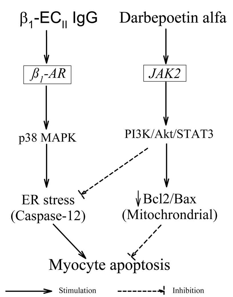
Dilated human cardiomyopathy is associated with suppression of the prosurvival phosphatidylinositol-3-kinase (PI3K)/Akt and STAT3 pathways. The present study was carried out to determine if restoration of the PI3K/Akt and STAT3 activity by darbepoetin alfa improved cardiac function or reduced cardiomyocyte apoptosis in rabbit autoimmune cardiomyopathy induced by a peptide corresponding to the second extracellular loop of the ss(1)-adrenergic receptor (ss(1)-EC(II)). We found that ss(1)-EC(II) immunization produced progressive LV dilation, systolic dysfunction and myocyte apoptosis as measured by TUNEL, single-stranded DNA antibody, and active caspase-3. These changes were associated with activation of p38 mitogen-activated protein kinase (MAPK), endoplasmic reticulum stress markers (GRP78 and CHOP), and increased cleavage of procaspase-12, as well as decreased phosphorylation of Akt and STAT3, and decreased Bcl2/Bax ratio. As expected, darbepoetin alfa treatment increased phosphorylation of Akt and STAT3. It also increased the myocardial expression of erythropoietin receptor which was reduced in the failing myocardium, and improved cardiac function in the ss(1)-EC(II)-immunized animals. The latter was associated with reductions of myocyte apoptosis and cleaved caspase-3, as well as reversal of increased phosphorylation of p38-MAPK, increased ER stress, and decline in Bcl2/Bax ratio. The anti-apoptotic effects of darbepoetin alfa via Akt and STAT activation were also demonstrated in cultured cardiomyocytes treated with the anti-ss(1)-EC(II) antibody. These effects of darbepoetin alfa in vitro were prevented by LY294002 and STAT3 peptide inhibitor. Thus, we conclude that darbepoetin alfa improves cardiac function and prevents progression of dilated cardiomyopathy probably by activating the PI3K/Akt and STAT3 pathways and reducing ER stress.


Figures

Figure 1
Figure 1
Changes in erythropoietin receptor (EpoR) expression in left ventricular myocardium of rabbits following β1-ECII immunization, darbepoetin alfa administration, or both. Panel A. Representative EpoR expression by immunohistochemistry. Panel B. EpoR protein expression by Western blots. N=6 animals in each group. Representative immunoblots for EpoR and GAPDH are shown on the left to the bar graph. Bar denote SEM. *P<0.05, compared to Sham immunization, † P<0.05, compared to β1-ECII immunization, and ‡ P<0.05, compared to darbepoetin alfa alone.
Figure 2
Figure 2
Effects of darbepoetin alfa on left ventricular myocardial phosphorylated Akt (p-Akt), phosphorylated STAT3 (p-STAT3), Bax and Bcl-2 in the Sham– and β1-ECII–immunized rabbits. Representative immunoblots are shown on the left to the bar graphs. p-Akt and p-STAT3 are expressed as ratios of total Akt and total STAT3, respectively, in the bar graphs. N=6 animals in each group. Bars denote SEM. *P<0.05, compared to Sham immunization, and † P<0.05, compared to β1-ECII immunization.
Figure 3
Figure 3
Effects of darbepoetin alfa on left ventricular myocardial p-38 MAP kinase and ERK1/2 in the Sham– and β1-ECII–immunized rabbits. Representative immunoblots are shown on the left to the bar graphs. Phosphorylated p38-MAPK (p-38) and phosphorylated ERK1/2 (p-ERK) were expressed as ratios of total p38-MAPK and total ERK1/2, respectively, in the bar graphs. N=6 animals in each group. Bars denote SEM. *P< 0.05, compared to Sham immunization. † P< 0.05 compared to β1-ECII immunization.
Figure 4
Figure 4
Effects of darbepoetin alfa on left ventricular myocardial GRP78, CHOP, ATF6, and caspase-12 in the Sham– and β1-ECII–immunized rabbits. Representative immunoblots are shown on the left to the bar graphs. N=6 animals in each group. Bar denote SEM. *P<0.05, compared to Sham immunization, and † P<0.05, compared to β1-ECII immunization.
Figure 5
Figure 5
Effects of β1-ECII IgG and darbepoetin alfa on cell apoptosis in cultured neonatal rat cardiomyocytes. Representative micrographs are given on the top panel showing the increase of TUNEL-positive cardiomyocytes (indicated by red arrows) after addition of β1-ECII IgG, and the cardioprotective effect of darbepoetin alfa in the β1-ECII IgG–treated cardiomyocytes (left panel). Cardiomyocytes are identified by the actinin and propidium iodide stains (right panel). The bottom panel shows the statistical difference in cardiomyocyte apoptosis among the groups. N=6 in each group. Bar denote SEM. *P<0.05, compared to Control, and † P<0.05, compared to β1-ECII IgG.
Figure 6
Figure 6
Effects of β1-ECII IgG, darbepoetin alfa, LY294002 and STAT3 inhibitor peptide on ER stress markers GRP78, CHOP, and cleaved caspase-12 (Figure 6A), and phosphorylated Akt (p-Akt) and phosphorylated STAT3 (p-STAT) (Figure 6B) in cultured neonatal rat cardiomyocytes. Representative immunoblots are shown below the corresponding bar graphs. N=6 in each group. Bar denote SEM. *P<0.05, compared to Control. † P<0.05, compared to β1-ECII IgG. ‡ P<0.05, compared to β1-ECII IgG plus darbepoetin alfa.
Figure 6
Figure 6
Effects of β1-ECII IgG, darbepoetin alfa, LY294002 and STAT3 inhibitor peptide on ER stress markers GRP78, CHOP, and cleaved caspase-12 (Figure 6A), and phosphorylated Akt (p-Akt) and phosphorylated STAT3 (p-STAT) (Figure 6B) in cultured neonatal rat cardiomyocytes. Representative immunoblots are shown below the corresponding bar graphs. N=6 in each group. Bar denote SEM. *P<0.05, compared to Control. † P<0.05, compared to β1-ECII IgG. ‡ P<0.05, compared to β1-ECII IgG plus darbepoetin alfa.
Figure 7
Figure 7
A schematic diagram showing the proposed mode of action of darbepoetin alfa on activation of the prosurvival JAK/PI3K/Akt pathway, and inhibition of the ER-stress–mediated cardiomyocyte apoptosis induced by β1-ECII IgG.

Similar articles

Cited by

References

    1. Alberts B, Johnson A, Lewis J, Raff M, Roberts K, Walter P. Intracellular compartments and protein sorting. 5th ed. New York: Garland Science; 2008. Molecular Biology of the Cell. Chapter 12; pp. 695–748.
    1. Kaufman RJ. Orchestrating the unfolded protein response in health and disease. J Clin Invest. 2002;110:1389–1398. - PMC - PubMed
    1. Glembotski CC. The role of the unfolded protein response in the heart. J Mol Cell Cardiol. 2008;44:453–459. - PMC - PubMed
    1. Okada K, Minamino T, Tsukamoto Y, Liao Y, Tsukamoto O, Takashima S, Hirata A, Fujita M, Nagamachi Y, Nakatani T, Yutani C, Ozawa K, Ogawa S, Tomoike H, Hori M, Kitakaze M. Prolonged endoplasmic reticulum stress in hypertrophic and failing heart after aortic constriction: possible contribution of endoplasmic reticulum stress to cardiac myocyte apoptosis. Circulation. 2004;110:705–712. - PubMed
    1. Tsukamoto O, Minamino T, Okada K-i, Shintani Y, Takashima S, Kato H, Liao Y, Okazaki H, Asai M, Hirata A, Fujita M, Asano Y, Yamazaki S, Asanuma H, Hori M, Kitakaze M. Depression of proteasome activities during the progression of cardiac dysfunction in pressure-overloaded heart of mice. Biochem Biophys Res Commun. 2006;340:1125–1133. - PubMed

Publication types

MeSH terms