. 2008 Oct;29(10):1986-93.
doi: 10.1093/carcin/bgn128. Epub 2008 Jun 26.

(-)-Epigallocatechin gallate causes internalization of the epidermal growth factor receptor in human colon cancer cells

Affiliations

(-)-Epigallocatechin gallate causes internalization of the epidermal growth factor receptor in human colon cancer cells

Seiji Adachi et al. Carcinogenesis. 2008 Oct.

Abstract

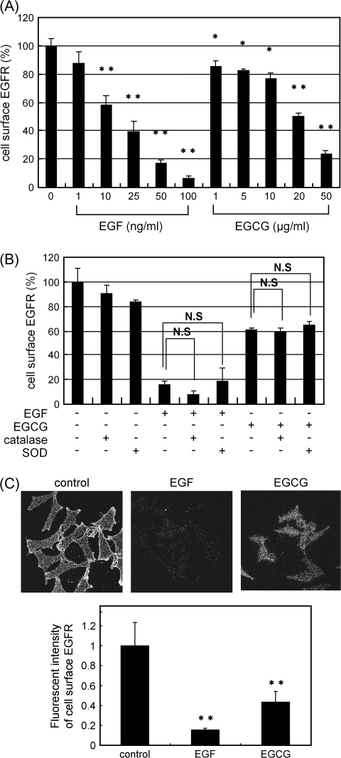
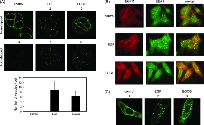
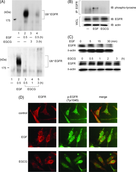
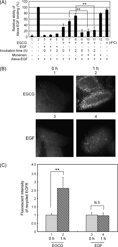
We recently found that the inhibitory effect of (-)-epigallocatechin gallate (EGCG) on epidermal growth factor (EGF) binding to the epidermal growth factor receptor (EGFR) is associated with alterations in lipid organization in the plasma membrane of colon cancer cells. Since changes in lipid organizations are thought to play a role in the trafficking of several membrane proteins, in this study we examined the effects of EGCG on cellular localization of the EGFR in SW480 cells. Treatment of the cells for 30 min with as little as 1 microg/ml of EGCG caused a decrease in cell surface-associated EGFRs and this was associated with internalization of EGFRs into endosomal vesicles. Similar effects were seen with a green fluorescent protein (GFP)-EGFR fusion protein. As expected, the EGFR protein was phosphorylated at tyrosine residues, ubiquitinated and partially degraded when the cells were treated with EGF, but treatment with EGCG caused none of these effects. The loss of EGFRs from the cell surface induced by treating the cells with EGF for 30 min persisted for at least 2 h. However, the loss of EGFRs from the cell surface induced by temporary exposure to EGCG was partially restored within 1-2 h. These studies provide the first evidence that EGCG can induce internalization of EGFRs into endosomes, which can recycle back to the cell surface. This sequestrating of inactivated EGFRs into endosomes may explain, at least in part, the ability of EGCG to inhibit activation of the EGFR and thereby exert anticancer effects.


Figures

Fig. 1.
Fig. 1.
Both EGF and EGCG induce decreases in cell surface-associated EGFR. (A) SW480 cells were first labeled for 15 min at 37°C with an anti-EGFR antibody that recognizes the extracellular domain of the EGFR. The cells were treated with EGF or EGCG at the indicated concentration for 30 min at 37°C. The amount of cell surface EGFR was then measured by ELISA (see Materials and Methods). The asterisks indicate a significant decrease (*P < 0.05, **P < 0.01, respectively) with respect to the control (first lane on left). (B) The cells were treated with EGF (100 ng/ml) or EGCG (20 μg/ml) with or without catalase (30 U/ml) or SOD (15 U/ml) for 30 min at 37°C. They were then fixed, exposed to the anti-EGFR antibody and the amount of cell surface-associated EGFR was measured by ELISA as described in (A). NS designates no significant difference between the indicated pairs. (C) The fluorescent intensities of cell surface EGFR was analyzed in untreated cells or cells treated with 100 ng/ml of EGF or 20 μg/ml of EGCG by fluorescence microscopy and the fluorescent intensity of cell surface EGFR was quantitated by the MetaMorph. Representative results from at least three independent experiments are shown. For additional details see Materials and Methods.
Fig. 2.
Fig. 2.
Both EGF and EGCG cause internalization of the EGFR. (A) SW480 cells were first labeled for 15 min at 37°C with an anti-EGFR antibody that recognizes the extracellular domain of the EGFR. They were then treated with EGF (100 ng/ml) or EGCG (20 μg/ml) for 30 min at 37°C, followed by fixation with paraformaldehyde. The fixed cells were then exposed to an Alexa 488-conjugated anti-mouse secondary antibody (green signal in upper panels 1–6), in the presence of 100 μg/ml of saponin to permeabilize the cells, to label the antibody–EGFR complex, and then examined by confocal microscopy (panels 1–3). In a second study (panels 4–6), the cells were treated as described above but they were exposed to acid-stripping buffer for 150 s before the fixation step (see Materials and Methods) to remove cell surface-associated EGFR antibody. Lower bar graph shows quantification data of the number of vesicles of the internalized EGFR shown in upper panels 4–6 (see details in Materials and Methods). (B) The cells were first labeled for 15 min at 37°C with an anti-EGFR antibody. They were then treated with EGF (100 ng/ml) or EGCG (20 μg/ml) for 30 min at 37°C and then fixed with paraformaldehyde. After permeabilization of the cells with 0.1% Triton X-100, the cells were exposed to anti-EEA1 antibody (1:100 dilution) for 1 h and then treated with Alexa 546-conjugated anti-mouse secondary antibody for EGFR (red signal) and Alexa 488-conjugated anti-mouse secondary antibody for EEA1 (green signal). They were then examined by confocal microscopy. (C) SW480 cells were transfected for 24 h with a plasmid encoding EGFR–GFP, prior to stimulation with the indicated compound. Thirty minutes after the addition of EGF (100 ng/ml) or EGCG (20 μg/ml) at 37°C, followed by fixation with paraformaldehyde and then examined by confocal microscopy. Representative results from at least three independent experiments are shown.
Fig. 3.
Fig. 3.
In contrast to the effects of EGF, EGCG does not cause ubiquitination and subsequent degradation of the EGFR. (A) Ubiquitination of the EGFR. The cells were pretreated with the proteasome inhibitor MG132 (10 μM) for 6 h and then exposed to EGF (100 ng/ml) for last 30 min or EGCG (20 μg/ml) for 0.5, 1 or 3 h at 37°C. Then, cell lysates (500 μg each) were prepared and incubated for 3 h at 4°C with an anti-EGFR antibody precoupled to anti-mouse IgG–agarose beads. The bound protein was then analyzed by western blotting with an anti-Ub antibody. (B) Phosphorylation at tyrosine residues of the EGFR. The cells were exposed to EGF (100 ng/ml) for 30 min or EGCG (20 μg/ml) for 30 min at 37°C. Each cell lysates were then prepared and incubated for 3 h at 4°C with an anti-EGFR antibody precoupled to anti-mouse IgG–agarose beads. The bound proteins were then analyzed by western blotting with an anti-phosphotyrosine antibody. The lower two panels indicate the corresponding whole cell lysates. (C) Rate of de novo proteolysis of the EGFR. The cells were treated with EGF (100 ng/ml) or EGCG (20 μg/ml) for the indicated time in the presence of cycloheximide (10 μg/ml) to block new protein synthesis. Protein extracts were prepared and examined by western blotting using an anti-EGFR antibody. An antibody to β-actin was used to control for protein loading. (D) Phosphorylation of the internalized EGFR on Tyr 1045 residues. The cells were first labeled for 15 min at 37°C with an anti-EGFR antibody. They were then treated with EGF (100 ng/ml) or EGCG (20 μg/ml) for 30 min at 37°C and then fixed with paraformaldehyde. After permeabilization of the cells with 0.1% Triton X-100, the cells were exposed to anti-phosphorylated EGFR (Tyr 1045) antibody for 1 h and then exposed to Alexa 546-conjugated anti-mouse secondary antibody for EGFR (red signal) and Alexa 488-conjugated anti-mouse secondary antibody for phosphorylated EGFR (Tyr 1045) (green signal). They were then examined by confocal microscopy. Representative results from at least three independent experiments are shown.
Fig. 4.
Fig. 4.
The EGFR that is internalized following transient treatment with EGCG can recycle back to the cell surface. (A) Quantification of cell surface EGFR using Alexa–EGF binding. The cells were treated with EGF (100 ng/ml) or EGCG (20 μg/ml) for 30 min at 37°C (lanes 1–12) or as a control they were incubated with EGCG at 4°C (lane 13). The medium was then changed and the cells were incubated in serum-free growth medium lacking EGF or EGCG at 37°C for 0, 1 or 2 h. They were then exposed to Alexa 488-conjugated EGF (100 ng/ml) for 1 h to label cell surface-associated EGFR. This was done at 4°C to prevent Alexa 488 EGF-induced internalization of the EGFR. As indicated in some assays, monensin (100 μM) was added at 15 min prior to the addition of EGF or EGCG to inhibit recycling of internalized vesicles. After the final incubation, the cells were washed with cold phosphate-buffered saline, harvested by the addition of trypsin and gentle scraping at 4°C and fixed with 3% paraformaldehyde for 10 min. They were then analyzed for cell surface-bound Alexa 488 EGF by flow cytometry. (B) Analysis of recycling of the EGFR by fluorescence microscopy. SW480 cells were first labeled for 15 min at 37°C with the anti-EGFR antibody that recognizes the extracellular domain of the EGFR for 15 min. They were then treated with EGF (100 ng/ml) or EGCG (20 μg/ml) for 30 min at 37°C. Any antibody remaining on the cell surface was then removed by treatment with the acid-stripping buffer for 150 s (see Materials and Methods). The cells were then not further incubated (panels 1 and 3) or they were incubated (panels 2 and 4) for 1 h at 37°C in serum-free growth medium without the addition of EGF or EGCG. They were then fixed with paraformaldehyde and exposed to the Alexa 488-conjugated anti-mouse secondary antibody in the absence of saponin and then examined by fluorescence microscopy. (C) Quantification of the amount of the EGFR that was recycled back to cell surface based on the fluorescent intensities in (B) which were analyzed by the MetaMorph. **P < 0.01, significant difference obtained by a comparison between indicated pairs. NS designates no significant difference obtained by a comparison between indicated pairs. Representative results from at least three independent experiments are shown.
Fig. 5.
Fig. 5.
A schematic diagram of the mechanism by which the EGF causes activation and internalization of the EGFR, and a hypothetical mechanism by which EGCG causes internalization of the EGFR. After EGF binds to EGFR molecules on the cell surface, the receptor undergoes dimerization and autophosphorylation at tyrosine residues, and this triggers EGFR-related downstream signaling. The EGFR is also ubiquitinated and internalized into early endosomes that are EEA1 positive, which become late endosomes, and eventually the receptors are degraded in lysosomes. By contrast, when cells are treated with EGCG, the receptor is not dimerized (26), autophosphorylated or ubiquitinated. However, EGFR molecules are also internalized into EEA1-positive early endosomes, perhaps because of EGCG-induced alterations in lipid organization (26), and they are not degraded at early time point. With time, these EGFR molecules can be recycled back to the cell surface.

References

    1. Masuda M, et al. Effects of epigallocatechin-3-gallate on growth, epidermal growth factor receptor signaling pathways, gene expression, and chemosensitivity in human head and neck squamous cell carcinoma cell lines. Clin. Cancer Res. 2001;7:4220–4229. - PubMed
    1. Pianetti S, et al. Green tea polyphenol epigallocatechin-3 gallate inhibits Her-2/neu signaling, proliferation, and transformed phenotype of breast cancer cells. Cancer Res. 2002;62:652–655. - PubMed
    1. Paschka AG, et al. Induction of apoptosis in prostate cancer cell lines by the green tea component, (-)-epigallocatechin-3-gallate. Cancer Lett. 1998;130:1–7. - PubMed
    1. Shimizu M, et al. (-)-Epigallocatechin gallate and polyphenon E inhibit growth and activation of the epidermal growth factor receptor and human epidermal growth factor receptor-2 signaling pathways in human colon cancer cells. Clin. Cancer Res. 2005;11:2735–2746. - PubMed
    1. Khan N, et al. Targeting multiple signaling pathways by green tea polyphenol (-)-epigallocatechin-3-gallate. Cancer Res. 2006;66:2500–2505. - PubMed

Publication types

MeSH terms