. 2008 Aug;36(13):4364-80.
doi: 10.1093/nar/gkn384. Epub 2008 Jun 27.

Mrd1p binds to pre-rRNA early during transcription independent of U3 snoRNA and is required for compaction of the pre-rRNA into small subunit processomes

Affiliations

Mrd1p binds to pre-rRNA early during transcription independent of U3 snoRNA and is required for compaction of the pre-rRNA into small subunit processomes

Asa Segerstolpe et al. Nucleic Acids Res. 2008 Aug.

Abstract

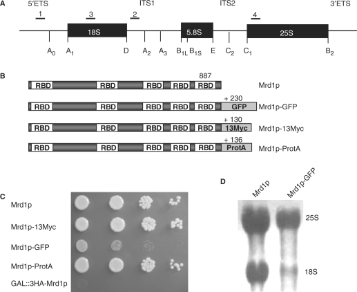
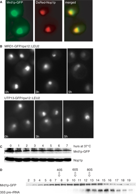
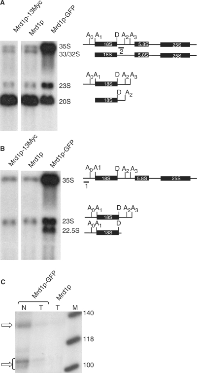
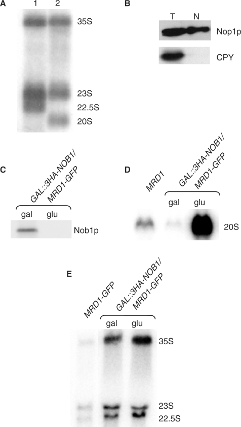
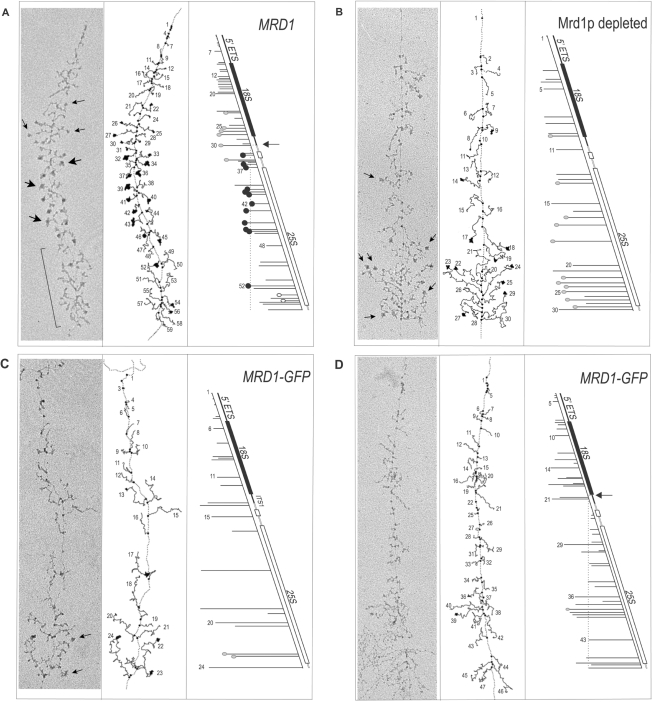
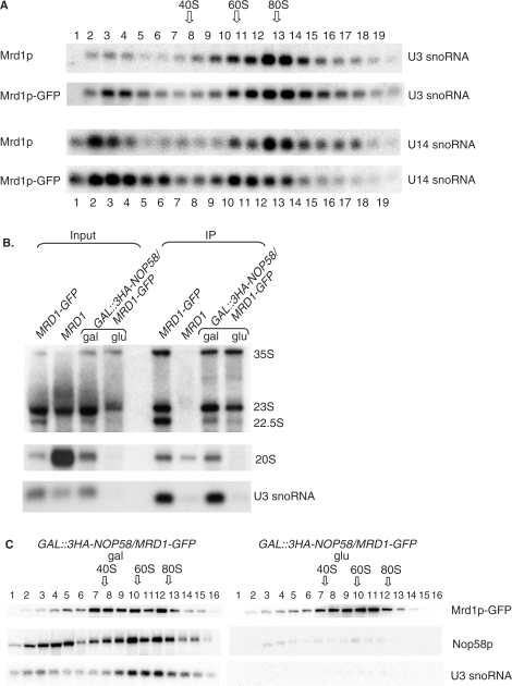
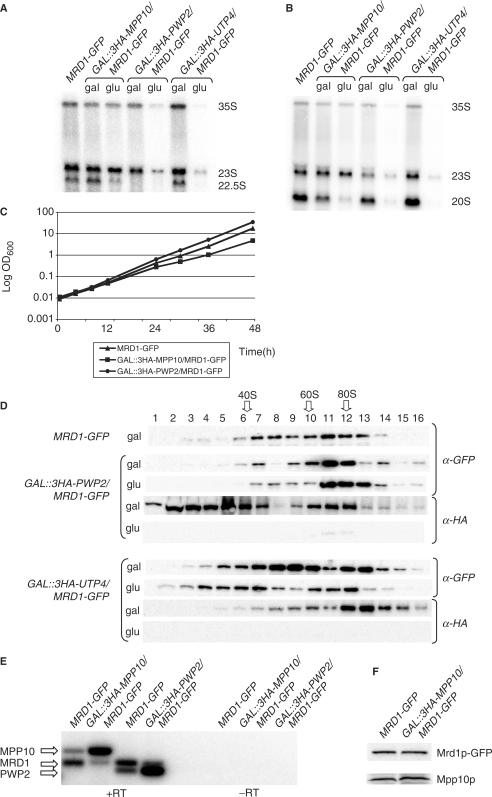
In Saccharomyces cerevisiae, synthesis of the small ribosomal subunit requires assembly of the 35S pre-rRNA into a 90S preribosomal complex. SnoRNAs, including U3 snoRNA, and many trans-acting proteins are required for the ordered assembly and function of the 90S preribosomal complex. Here, we show that the conserved protein Mrd1p binds to the pre-rRNA early during transcription and is required for compaction of the pre-18S rRNA into SSU processome particles. We have exploited the fact that an Mrd1p-GFP fusion protein is incorporated into the 90S preribosomal complex, where it acts as a partial loss-of-function mutation. When associated with the pre-rRNA, Mrd1p-GFP functionally interacts with the essential Pwp2, Mpp10 and U3 snoRNP subcomplexes that are functionally interconnected in the 90S preribosomal complex. The fusion protein can partially support 90S preribosome-mediated cleavages at the A(0)-A(2) sites. At the same time, on a substantial fraction of transcripts, the composition and/or structure of the 90S preribosomal complex is perturbed by the fusion protein in such a way that cleavage of the 35S pre-rRNA is either blocked or shifted to aberrant sites. These results show that Mrd1p is required for establishing productive structures within the 90S preribosomal complex.


Figures

Figure 1.
Figure 1.
The Mrd1p-GFP fusion protein acts as a partial loss-of-function mutation of Mrd1p. (A) Schematic representation of the 35S pre-rRNA. A0 to E represent cleavages during processing. The numbers 1–4 show the positions of the oligonucleotides used as probes in northern blot hybridizations. (B) Schematic representation of Mrd1p and modified versions of Mrd1p used in this study. Positions of the five RBDs in Mrd1p and the lengths of Mrd1p in amino acid residues and the different tags are shown. (C) Growth on YPD of strains containing Mrd1p or the different versions of Mrd1p. The doubling time was ∼110 min for Mrd1p, 106 min for Mrd1p-13Myc, 300 min for Mrd1p-GFP and 100 min for Mrd1p-ProtA. Depletion of Mrd1p (GAL::3HA-Mrd1p) is not compatible with growth (bottom row). (D) RNA was extracted from cells containing Mrd1p or Mrd1p-GFP, subjected to electrophoresis in 1% agarose/formaldehyde gels and transferred to a membrane. The 25S and 18S rRNAs were visualized by methylene blue staining.
Figure 2.
Figure 2.
Mrd1p-GFP is located primarily in the nucleolus and this localization is dependent on transcription by RNA polymerase I. (A) Mrd1p-GFP and DsRed-Nop1p colocalized in the nucleolus. The Mrd1p-GFP strain was transformed with pUN100-DsRed-NOP1 and grown in synthetic media lacking leucine and analyzed with fluorescence microscopy. (B) Localization of Mrd1p-GFP and Utp13p–GFP by fluorescent microscopy after shut off of RNA polymerase I transcription. Cells containing Mrd1p-GFP or Utp13p–GFP together with a temperature sensitive RNA polymerase I allele (rpa12::LEU2) were grown at permissive temperature (0 h) and shifted to 37°C for 3 h and 5 h. (C) Stability of Mrd1p-GFP analyzed by western blotting. Cells containing Mrd1p-GFP and a temperature-sensitive RNA polymerase I allele (rpa12::LEU2) were grown at permissive temperature (0 h) and shifted to 37°C for 1–7 h. Nop1p acted as a loading control. (D) Sucrose gradient analyses of extracts from cells with Mrd1p-GFP. Mrd1p-GFP was identified by western blot analyses of each fraction. The 35S pre-rRNA was identified by northern blot analyses using probe 1. The positions of 40S, 60S and 80S are indicated, as determined from methylene blue staining of the RNA in each fraction.
Figure 3.
Figure 3.
Mrd1p-GFP partially inhibits the A0–A2 cleavages and induces aberrant processing products with 3′-ends located in a narrow region just downstream of the D site. (A and B) Northern hybridizations using probes specific for pre-rRNA species. RNA was extracted from wild-type cells (Mrd1p), Mrd1p-13Myc and Mrd1p-GFP cells and separated on 1% agarose/formaldehyde gels. Schematic representations of identified pre-rRNA species and probes used are indicated to the right of each panel. Probe 2 was used in (A), and probe 1 was used in (B). (C) RNase protection analyses using a 164-nt long fragment covering the 18S rRNA-ITS1 border. RNA was extracted from purified nuclei and from cells expressing Mrd1p-GFP or from cells expressing Mrd1p. The probe extended from a position 100-nt upstream of the D cleavage site to a position located 64-nt downstream of the D site. Protected RNAs, indicated by arrows, were analyzed in 8% polyacrylamide/urea gels. N, nuclear RNA; T, total cellular RNA; M, size markers.
Figure 4.
Figure 4.
The aberrant cleavages producing 22.5S rRNA take place in the cell nucleus and are not dependent on Nob1p. (A) RNA isolated from cell nuclei purified from Mrd1p-GFP cells was analyzed by northern hybridization. The probes used were as follows: lane 1, probe 1; lane 2, probe 2. (B) Western blot analyses of Nop1p and CPY in extracts from total cells (T) or from purified nuclei (N). (C) Western blot analyses of GAL::3HA-NOB1/MRD1-GFP, grown in YPG (gal) or YPD (glu) for 15 h. (D) Northern hybridization using probe 2 of total RNA from wild-type (Mrd1p) and GAL::3HA-NOB1/MRD1-GFP cells, grown in YPG (gal) or YPD (glu). The 20S pre-rRNA is shown. (E) Northern hybridizations using probe 1 of total RNA isolated from MRD1-GFP, GAL::3HA-NOB1/MRD1-GFP grown in YPG (gal) or YPD (glu).
Figure 5.
Figure 5.
Mrd1p-GFP disturbs the cotranscriptional assembly of 90S preribosomal complexes. (A) Representative rRNA gene from control cells (Mrd1p) and an interpretive tracing of the gene, with small arrows indicating examples of small terminal knobs (thought to contain U3 snoRNP), and larger arrows indicating examples of SSU processomes. Bracketed region indicates cleaved transcript region. A transcript map of the gene is shown on the right. Transcripts are linearized and shown at the appropriate position along the gene. The gene is drawn on a slope so that the transcript sequences are approximately aligned in sequence, with 5′-termini to the left. Small (gray) and large (black) terminal particles are shown. For reference, the dotted line extrapolates to the site of A2 cleavage, which is indicated by an arrow on the gene map. (B) The rRNA gene from cells in which Mrd1p was depleted for 20 h. Formation of 5′ particles (arrows) occurred, but SSU processomes were not formed. (C) The rRNA gene from Mrd1p-GFP cells showing little cotranscriptional A2 cleavage of transcripts. (D) The rRNA gene from Mrd1p-GFP cells showing an unusual type of cotranscriptional cleavage. For reference, dotted line and arrow in the panel indicate the approximate position of the 3′-ends in the 22.5S rRNA (see text).
Figure 6.
Figure 6.
Mrd1p-GFP is compatible with the presence of U3 and U14 snoRNAs in 90S preribosomal complexes and U3 snoRNP is required for induction of the aberrant 22.5S rRNA, but not for Mrd1p-GFP association with 35S pre-rRNA and 23S rRNA. (A) Northern hybridizations using U3 or U14 snoRNA probes. Extracts from wt (Mrd1p) or Mrd1p-GFP were subjected to sucrose gradient centrifugation. RNA was extracted from each fraction and separated in 1.2% agarose/formaldehyde gels. The positions of 40S, 60S and 80S are indicated as determined from methylene blue staining of RNA in each fraction. (B) Extracts from cells with Mrd1p-GFP and Mrd1p grown in YPD and GAL::3HA-NOP58/MRD1-GFP grown in YPG (gal) or YPD (glu) for 16 h were immunoprecipitated with anti-GFP antibodies. RNA in the extracts (Input, 1/30 of total extract) and coimmunoprecipitated RNA (IP) was size separated on a 1.5% agarose gel and probed with probe 1, probe 2 and U3 probe. (C) Distribution of Mrd1p-GFP, 3HA-Nop58p and U3 snoRNA in 10–50% sucrose gradients. Extracts from GAL::3HA-NOP58/MRD1-GFP cells grown in YPG (gal) or YPD (glu) for 17 h were analyzed by western blotting or northern blotting with the U3 probe. The positions of 40S, 60S and 80S are indicated.
Figure 7.
Figure 7.
Pwp2p and Mpp10p influence the function of Mrd1p-GFP. (A) Northern hybridization using probe 1. Total RNA was extracted from Mrd1p-GFP cells and from cells with GAL::3HA-MPP10/MRD1-GFP, GAL::3HA-PWP2/MRD1-GFP and GAL::3HA-UTP4/MRD1-GFP grown in YPG (gal) or YPD (glu). The positions of pre-rRNA species are indicated to the right. The ratio between the hybridization signals for 22.5S and 23S were: 0.6 for MRD-GFP, 0.8 for MPP10 (gal), 0.2 for PWP2 (gal) and 0.6 for UTP4 (gal). (B) Northern hybridization using probe 2. RNA was extracted from cells as in (A). The ratio between the hybridization signals for 20S and 23S were:1.3 for MRD-GFP, 0.9 for MPP10 (gal), 1.7 for PWP2 (gal) and 1.6 for UTP4 (gal). (C) Comparison of the growth rate of MRD1-GFP, GAL::3HA-MPP10/MRD1-GFP and GAL::3HA-PWP2/MRD1-GFP cells grown in YPG media for the time indicated. Cells were kept in exponential growth throughout the experiment by dilution in prewarmed medium. (D) Western blot analyses of Mrd1p-GFP, 3HA-Pwp2p and 3HA-Utp4p in fractions from sucrose gradients of cells containing MRD1-GFP, GAL::3HA-PWP2/MRD1-GFP and GAL::3HA-UTP4/MRD1-GFP. Cells were grown in YPG (gal) or YPD (glu) for 16 h. The positions of 40S, 60S and 80S are indicated. (E) Relative mRNA levels measured by RT–PCR. RNA was extracted from cells grown in galactose medium. These cells contained either the MRD1-GFP gene and all other genes expressed from their wild-type promoters (MRD1-GFP), or the MPP10 gene expressed from the GAL1 promoter and all other genes, including MRD1-GFP expressed from their wild-type promoters (GAL::3HA-MPP10/MRD1-GFP), or the PWP2 gene expressed from the GAL1 promoter and all other genes, including MRD1-GFP expressed from their wild-type promoters (GAL::3HA-PWP2/MRD1-GFP). PCR products corresponding to the different mRNAs are indicated. (F) Western blot of Mrd1p-GFP and Mpp10p in PLY129 and PLY339. The strains were grown in galactose medium. Proteins were extracted from the same number of cells and analyzed by western blotting, using an anti-GFP antibody and anti-Mpp10p antibodies.

Similar articles

Cited by

References

    1. Venema J, Tollervey D. Ribosome synthesis in Saccharomyces cerevisiae. Annu. Rev. Genet. 1999;33:261–311. - PubMed
    1. Udem SR, Warner JR. Ribosomal RNA synthesis in Saccharomyces cerevisiae. J. Mol. Biol. 1972;65:227–242. - PubMed
    1. Trapman J, Retél J, Planta RJ. Ribosomal precursor particles from yeast. Exp. Cell Res. 1975;90:95–104. - PubMed
    1. Osheim YN, French SL, Keck KM, Champion EA, Spasov K, Dragon F, Baserga SJ, Beyer AL. Pre-18S ribosomal RNA is structurally compacted into the SSU processome prior to being cleaved from nascent transcripts in Saccharomyces cerevisiae. Mol. Cell. 2004;16:943–954. - PubMed
    1. Dragon F, Gallagher JE, Compagnone-Post PA, Mitchell BM, Porwancher KA, Wehner KA, Wormsley S, Settlage RE, Shabanowitz J, Osheim Y, et al. A large nucleolar U3 ribonucleoprotein required for 18S ribosomal RNA biogenesis. Nature. 2002;417:967–970. - PMC - PubMed

Publication types

MeSH terms