. 2008 Aug;295(2):H874-82.
doi: 10.1152/ajpheart.01189.2007. Epub 2008 Jun 27.

Intramitochondrial signaling: interactions among mitoKATP, PKCepsilon, ROS, and MPT

Affiliations

Intramitochondrial signaling: interactions among mitoKATP, PKCepsilon, ROS, and MPT

Alexandre D T Costa et al. Am J Physiol Heart Circ Physiol. 2008 Aug.

Abstract

Activation of protein kinase Cepsilon (PKCepsilon), opening of mitochondrial ATP-sensitive K(+) channels (mitoK(ATP)), and increased mitochondrial reactive oxygen species (ROS) are key events in the signaling that underlies cardioprotection. We showed previously that mitoK(ATP) is opened by activation of a mitochondrial PKCepsilon, designated PKCepsilon1, that is closely associated with mitoK(ATP). mitoK(ATP) opening then causes an increase in ROS production by complex I of the respiratory chain. This ROS activates a second pool of PKCepsilon, designated PKCepsilon2, which inhibits the mitochondrial permeability transition (MPT). In the present study, we measured mitoK(ATP)-dependent changes in mitochondrial matrix volume to further investigate the relationships among PKCepsilon, mitoK(ATP), ROS, and MPT. We present evidence that 1) mitoK(ATP) can be opened by H(2)O(2) and nitric oxide (NO) and that these effects are mediated by PKCepsilon1 and not by direct actions on mitoK(ATP), 2) superoxide has no effect on mitoK(ATP) opening, 3) exogenous H(2)O(2) or NO also inhibits MPT opening, and both compounds do so independently of mitoK(ATP) activity via activation of PKCepsilon2, 4) mitoK(ATP) opening induced by PKG, phorbol ester, or diazoxide is not mediated by ROS, and 5) mitoK(ATP)-generated ROS activates PKCepsilon1 and induces phosphorylation-dependent mitoK(ATP) opening in vitro and in vivo. Thus mitoK(ATP)-dependent mitoK(ATP) opening constitutes a positive feedback loop capable of maintaining the channel open after the stimulus is no longer present. This feedback pathway may be responsible for the lasting protective effect of preconditioning, colloquially known as the memory effect.


Figures

Fig. 1.
Fig. 1.
PKCɛ-mediated mitochondrial ATP-sensitive K+ channel (mitoKATP) opening. Changes in mitochondrial matrix volume (V) are plotted vs. time. Rat heart mitochondria (0.1 mg/ml) were suspended in assay medium described in methods. H2O2 (2 μM) or ψɛ receptor for activated C kinase (RACK) (0.5 μM) was added to medium in the presence of ATP (0.2 mM) ∼1 s after the mitochondria. Other additions to the assay medium were 5-hydroxydecanoate (5-HD, 0.3 mM) and ɛV1-2 (0.5 μM). Traces are representative of at least 5 independent experiments.
Fig. 2.
Fig. 2.
H2O2-induced mitoKATP opening is blocked by PKCɛ inhibitors. Shown are the effects of several compounds on mitoKATP opening induced by H2O2 plotted as volume change (%). Indicated additions to the assay were ATP (0.2 mM), H2O2 (2 μM), 5-HD (0.3 mM), glibenclamide (glib, 10 μM), Ro-318220 (Ro, 0.5 μM), ɛV1-2 (0.5 μM), PKCɛ scrambled peptide (scr, 1 μM), δV1-1 (0.2 μM), genistein (gen, 5 μM), and cyclosporin A (CsA, 1 μM). Data are means ± SD of at least 4 independent experiments.
Fig. 3.
Fig. 3.
H2O2, but not superoxide, opens mitoKATP via PKCɛ. Shown are the effects of xanthine oxidase (XOx) + hypoxanthine on mitoKATP activity, plotted as volume change (%). Rat heart mitochondria (0.1 mg/ml) were suspended in assay medium containing ATP (0.2 mM) and hypoxanthine (hypo, 0.2 μM). XOx (6 or 60 U/ml) was added to medium ∼1 s after mitochondria. As indicated, medium also contained catalase (cata, 10 U/ml), superoxide dismutase (SOD, 30 U), 5-HD (0.3 mM), and ɛV1-2 (0.5 μM). Data are means ± SD of at least 4 independent experiments.
Fig. 4.
Fig. 4.
Nitric oxide (NO) opens mitoKATP via PKCɛ. Shown are the effects of the NO donor S-nitroso-N-acetylpenicillamine (SNAP) on mitoKATP activity, plotted as volume change (%). Rat heart mitochondria (0.1 mg/ml) were suspended in assay medium containing ATP (0.2 mM). SNAP (10 mM) was added 2 min before mitochondria to allow generation of NO. As indicated, medium also contained 5-HD (0.3 mM), ɛV1-2 (0.5 μM), and catalase (10 U/ml). Columns on right demonstrate the results of experiments with mitoplasts lacking the mitochondrial outer membrane (MOM). N-(2-mercaptopropionyl)glycine (MPG, 0.3 mM) and protein phosphatase 2A (PP2A, 11 ng/ml) blocked mitoKATP opening by SNAP in mitoplasts. Data are means ± SD of at least 4 independent experiments.
Fig. 5.
Fig. 5.
The MOM is essential for PKG-induced, but not PKCɛ-induced, mitoKATP activity. Shown are the effects of PKG or PMA, and PP2A, on the matrix volume of heart mitochondria or mitoplasts. Data are plotted as volume change (%). Indicated additions to the assay were PKG (25 ng/ml), PMA (0.2 μM), and PP2A (11 ng/ml). Data are means ± SD of at least 4 independent experiments.
Fig. 6.
Fig. 6.
mitoKATP opening by diazoxide, PKG, or PMA does not require reactive oxygen species (ROS). Shown are the effects of MPG on diazoxide (Dzx)-, PMA-, or PKG/cGMP-induced mitoKATP opening, plotted as volume change (%). Rat heart mitochondria (0.1 mg/ml) were suspended in assay medium as described in methods. Additions to the assay were ATP (0.2 mM), Dzx (30 μM), PKG (25 ng/ml), cGMP (10 μM), PMA (0.2 μM), 5-HD (0.3 mM), chelerythrine (chel, 0.1 μM), MPG (0.3 mM), and KT-5823 (KT, 0.5 μM). Data are means ± SD of at least 4 independent experiments.
Fig. 7.
Fig. 7.
Phosphorylation-dependent mitoKATP-induced mitoKATP opening in vitro. Shown are the effects of preincubating heart mitoplasts (0.3 mg) with ATP (0.2 mM) + Dzx (30 μM), plotted as volume change (%). Preincubations were carried out in assay medium containing phosphatase inhibitors and, where indicated, ɛV1-2 (0.5 μM) or MPG (0.3 mM, 0.5%) at 30°C. After 3 min, the mitoplasts were washed and diluted 100-fold into the assay medium in order to avoid effects of Dzx, ɛV1-2, or MPG during the assay. Indicated additions to the assay were ATP (0.2 mM), Dzx (30 μM), and PP2A (11 ng/ml). Data are means ± SD of at least 3 independent experiments.
Fig. 8.
Fig. 8.
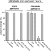
Phosphorylation-dependent persistence of mitoKATP opening in diazoxide-perfused hearts. Shown are the effects of sham perfusion or Dzx perfusion on mitoKATP activity in mitoplasts isolated from perfused rat hearts. mitoKATP activity is plotted as volume change (%). Indicated additions to the assay were ATP (0.2 mM), Dzx (30 μM), and PP2A (11 ng/ml). Data are means ± SD of at least 3 independent experiments.
Fig. 9.
Fig. 9.
NO inhibits mitochondrial permeability transition (MPT) via PKCɛ and independently of mitoKATP. Shown are the effects of Dzx or the NO donor SNAP on MPT-induced swelling, expressed as MPT inhibition (%). Synchronized MPT opening in rat heart mitochondria (0.1 mg/ml) was elicited by Ca2+ and the uncoupler CCCP, as described in methods. Rates of matrix swelling in the presence and absence of 1 μM CsA were taken as 0% and 100%, respectively. SNAP (10 mM) was added 2 min before mitochondria to allow generation of NO. 5-HD (0.3 mM) and ɛV1-2 (0.5 μM) were added immediately before mitochondria. Data are means ± SD of at least 3 independent experiments.
Fig. 10.
Fig. 10.
The intramitochondrial signaling pathways. There are 3 distinct ways of opening mitoKATP and initiating the signaling sequence described. 1) Direct mitoKATP opening by KATP channel openers (KCO) has been demonstrated in mitochondria and in liposomes containing reconstituted mitoKATP (22). 2) Indirect mitoKATP opening by activation of PKCɛ1 was demonstrated by the effects of the PKCɛ-specific peptide agonist ψɛRACK, PMA, H2O2, and NO. That these agents were acting via PKCɛ1 was verified by the finding that the PKCɛ-specific binding antagonist ɛV1-2 blocked all 4 modes of PKCɛ activation of mitoKATP but did not block mitoKATP opening by diazoxide (Ref. and present study). PKCɛ1 effect requires phosphorylation, perhaps of mitoKATP itself. Thus, when given access in mitoplasts to the mitochondrial inner membrane (MIM), PP2A prevented mitoKATP-dependent swelling induced by PKCɛ agonists. 3) Physiological mitoKATP opening by signals arriving at the MOM from the cytosol, such as PKG (6). PKG + cGMP open mitoKATP by phosphorylating a MOM receptor (labeled R1). Thus PKG-dependent mitoKATP opening is blocked either if PP2A is added to the assay or if the MOM is removed. Phosphorylation of R1 leads by an unknown mechanism to activation of PKCɛ1 and opening of mitoKATP. Once mitoKATP is opened, the increase in K+ uptake leads to increased matrix volume (ΔV), which is the basis of the light scattering assay for mitoKATP activity (8). More K+ than phosphate will be taken up into the matrix, because the cytosolic concentration of K+ is much higher than that of phosphate. This imbalance leads to matrix alkalinization (8). Matrix alkalinization, in turn, inhibits complex I, leading to increased production of superoxide and its products, H2O2 and hydroxyl anion radical (1). The increase in ROS plays 2 roles. It activates a second PKCɛ, PKCɛ2, that then inhibits the MPT in a phosphorylation-dependent reaction (7). We hypothesize that this effect is the primary means by which preconditioning and ischemic postconditioning prevent cardiac cell death. The increase in ROS also activates PKCɛ1, which is bypassed when KCOs are administered to the heart, to cause a persistent phosphorylation-dependent open state of mitoKATP (present study). We hypothesize that this positive feedback loop for mitoKATP opening is the mechanism of memory, which is seen with all preconditioning triggers (15, 47).

References

    1. Andrukhiv A, Costa AD, West IC, Garlid KD. Opening mitoKATP increases superoxide generation from complex I of the electron transport chain. Am J Physiol Heart Circ Physiol 291: H2067–H2074, 2006. - PubMed
    1. Argaud L, Gateau-Roesch O, Chalabreysse L, Gomez L, Loufouat J, Thivolet-Bejui F, Robert D, Ovize M. Preconditioning delays Ca2+-induced mitochondrial permeability transition. Cardiovasc Res 61: 115–122, 2004. - PubMed
    1. Argaud L, Gateau-Roesch O, Raisky O, Loufouat J, Robert D, Ovize M. Postconditioning inhibits mitochondrial permeability transition. Circulation 111: 194–197, 2005. - PubMed
    1. Beavis AD, Brannan RD, Garlid KD. Swelling and contraction of the mitochondrial matrix. I. A structural interpretation of the relationship between light scattering and matrix volume. J Biol Chem 260: 13424–13433, 1985. - PubMed
    1. Brookes PS, Salinas EP, Darley-Usmar K, Eiserich JP, Freeman BA, Darley-Usmar VM, Anderson PG. Concentration-dependent effects of nitric oxide on mitochondrial permeability transition and cytochrome c release. J Biol Chem 275: 20474–20479, 2000. - PubMed

Publication types

MeSH terms

LinkOut - more resources