. 2008 Aug;295(2):H883-9.
doi: 10.1152/ajpheart.00466.2008. Epub 2008 Jun 27.

Chemical ablation of the Purkinje system causes early termination and activation rate slowing of long-duration ventricular fibrillation in dogs

Affiliations

Chemical ablation of the Purkinje system causes early termination and activation rate slowing of long-duration ventricular fibrillation in dogs

Derek J Dosdall et al. Am J Physiol Heart Circ Physiol. 2008 Aug.

Abstract

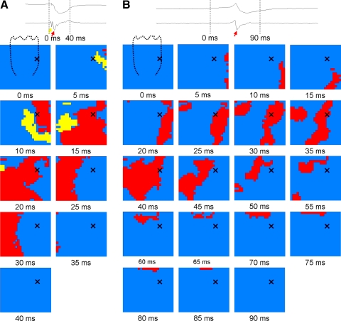
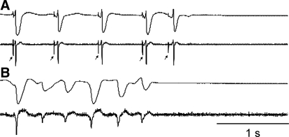
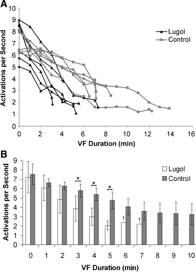
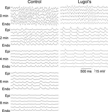
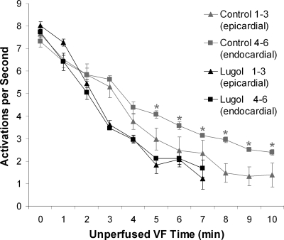
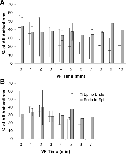
Endocardial mapping has suggested that Purkinje fibers may play a role in the maintenance of long-duration ventricular fibrillation (LDVF). To determine the influence of Purkinje fibers on LDVF, we chemically ablated the Purkinje system with Lugol solution and recorded endocardial and transmural activation during LDVF. Dog hearts were isolated and perfused, and the ventricular endocardium was exposed and treated with Lugol solution (n = 6) or normal Tyrode solution as a control (n = 6). The left anterior papillary muscle endocardium was mapped with a 504-electrode (21 x 24) plaque with electrodes spaced 1 mm apart. Transmural activation was recorded with a six-electrode plunge needle on each side of the plaque. Ventricular fibrillation (VF) was induced, and perfusion was halted. LDVF spontaneously terminated sooner in Lugol-ablated hearts than in control hearts (4.9 +/- 1.5 vs. 9.2 +/- 3.2 min, P = 0.01). After termination of VF, both the control and Lugol hearts were typically excitable, but only short episodes of VF could be reinduced. Endocardial activation rates were similar during the first 2 min of LDVF for Lugol-ablated and control hearts but were significantly slower in Lugol hearts by 3 min. In control hearts, the endocardium activated more rapidly than the epicardium after 4 min of LDVF with wave fronts propagating most often from the endocardium to epicardium. No difference in transmural activation rate or wave front direction was observed in Lugol hearts. Ablation of the subendocardium hastens VF spontaneous termination and alters VF activation sequences, suggesting that Purkinje fibers are important in the maintenance of LDVF.


Figures

Fig. 1.
Fig. 1.
Purkinje activations recorded during a paced beat before (A) but not after (B) Lugol ablation. A: the voltage electrogram (top trace) and the temporal derivative (bottom trace) at the top of the panel were recorded from the location indicated by an X on the plaque below. The arrows in the electrograms denote Purkinje (yellow arrow) and working myocardial (red arrows) activations. The plaque below represents the 504 electrodes. The dashed line in the frame of 0 ms represents the approximate location of the anterior papillary muscle. The timing of the frame (in ms) is shown below each panel. Electrodes with derivatives >−0.2 V/s at the time frame shown are inactive (blue). Purkinje activations (yellow) and activations of the working myocardium (red) are shown when the current frame electrogram signal has a derivative of ≤−0.2 V/s. Before Lugol ablation, activation from a paced beat first appears in the Purkinje system (yellow) and then in the working myocardium (red). The wave front activates the entire plaque in less than 40 ms. B: a paced beat in the same heart following Lugol ablation. The activation proceeds from the area to the right of the papillary muscle to the area to the left of the papillary muscle, with the papillary muscle activating last. The wave front takes nearly 90 ms to activate the entire mapped region.
Fig. 2.
Fig. 2.
Electrical activations were recorded until long-duration ventricular fibrillation (LDVF) abruptly terminated. A: an electrogram (top) and its temporal derivative (bottom) from a plaque in a control heart demonstrate the termination of LDVF. Purkinje activations were recorded in control hearts until the termination of LDVF. B: an electrogram (top) and its temporal derivative (bottom) in a Lugol-ablated heart show spontaneous termination of LDVF. No Purkinje activations were recorded in Lugol-treated hearts.
Fig. 3.
Fig. 3.
Activation rates of plaque electrodes and ventricular fibrillation (VF) termination times. A: mean activation rates recorded by the plaque electrodes and VF termination times as determined by the plaque and plunge needle recordings are shown for each Lugol-ablated (red) and control (blue) heart. B: average activation rates (mean with bars showing standard deviation) for all Lugol-ablated (red) together and all control (blue) hearts in which VF persisted are shown. VF durations with P < 0.05 by an unpaired t-test are marked with an asterisk. Statistics were not performed for minutes 6 and 7 (marked with †) because VF persisted in only 1 Lugol-ablated heart at this time. VF did not last more than 7 min in any Lugol-ablated heart.
Fig. 4.
Fig. 4.
Recordings from the 6 electrodes of a plunge needle in a control and a Lugol-ablated heart every 2 min during VF. As VF progresses, an activation rate gradient develops in the control animals in which the endocardium (Endo) activates more rapidly than the epicardium (Epi). In the Lugol-ablated heart, the endocardium does not activate more rapidly than the epicardium. VF terminated at 8.25 min in the control heart and at 4.5 min in the Lugol-treated heart shown in this figure.
Fig. 5.
Fig. 5.
Mean activation rate of the 3 most endocardial and the 3 most epicardial plunge needle electrodes during VF. An activation rate gradient developed after 4 min of VF in the control hearts with the endocardial electrodes activating significantly faster than the epicardial electrodes but did not develop in the Lugol-ablated hearts. Mean activation rate with the standard deviation is shown. Statistically different values are denoted with asterisks.
Fig. 6.
Fig. 6.
Mean direction of the VF wave fronts along the plunge needles. Wavefronts in control hearts propagated significantly more often from the endocardium toward the epicardium than in the opposite direction (A), whereas there was no preferential direction of propagation in Lugol-ablated hearts (B).

Similar articles

Cited by

References

    1. AHA Special Report. Position of the American Heart Association on research animal use. Circulation 71: 849A–850A, 1985. - PubMed
    1. Allison JS, Qin H, Dosdall DJ, Huang J, Newton JC, Allred JD, Smith WM, Ideker RE. The transmural activation sequence in porcine and canine left ventricle is markedly different during long-duration ventricular fibrillation. J Cardiovasc Electrophysiol 18: 1306–1312, 2007. - PubMed
    1. Antzelevitch C, Sicouri S, Litovsky SH, Lukas A, Krishnan SC, Di Diego JM, Gintant GA, Liu DW. Heterogeneity within the ventricular wall: electrophysiology and pharmacology of epicardial, endocardial, and M cells. Circ Res 69: 1427–1449, 1991. - PubMed
    1. Arnar DO, Bullinga JR, Martins JB. Role of the Purkinje system in spontaneous ventricular tachycardia during acute ischemia in a canine model. Circulation 96: 2421–2429, 1997. - PubMed
    1. Arnar DO, Martins JB. Purkinje involvement in arrhythmias after coronary artery reperfusion. Am J Physiol Heart Circ Physiol 282: H1189–H1196, 2002. - PubMed

Publication types

LinkOut - more resources