

Review
doi: 10.2337/db07-1769.
Protein kinase C function in muscle, liver, and beta-cells and its therapeutic implications for type 2 diabetes
Affiliations
- PMID: 18586909
- PMCID: PMC2453608
- DOI: 10.2337/db07-1769
Item in Clipboard
Review
Protein kinase C function in muscle, liver, and beta-cells and its therapeutic implications for type 2 diabetes
Diabetes.
2008 Jul.
No abstract available
Figures
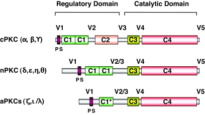
The PKC family of lipid-activated protein kinases. PKC isoforms contain constant regions (C1–4) and variable regions (V1–5) and can be divided into three subgroups. cPKCs are activated in the presence of calcium, which binds to the C2 domain, and DAG, which binds to the C1 domains. nPKCs lack C2 domains and are Ca2+-independent but still require DAG for full activation. aPKCs possess only one nonfunctional C1 domain (C1*) and no C2 domain and are both Ca2+- and DAG-independent. The C3 regions (ATP-binding) and C4 regions (protein substrate binding) are highly conserved between isoforms. In each case, the pseudosubstrate (PS) sequences, found in the V1 variable region, interfere with the catalytic domains to inhibit substrate phosphorylation until conformational changes induced by activators allow full activation.
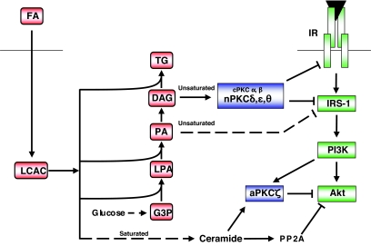
Lipid oversupply leads to the generation of distinct intracellular mediators of insulin resistance. Fatty acids entering the cell are activated by the formation of LCAC. Saturated fatty acid favors ceramide accumulation due to the requirement for palmitate during de novo synthesis, which in turn leads to the inhibition of Akt, in part due to aPKCζ action. In contrast, DAG species derived from unsaturated fatty acids favor nPKC activation and inhibition at the level of the insulin receptor (IR), or IRS-1. In addition, another unsaturated fatty acid–derived species, dilinoleoyl-phosphatidic acid (PA), can reduce IRS-1 tyrosine phosphorylation in a PKC-independent manner (49). G3P, glycerol 3-phosphate; LPA, lysophosphatidic acid; TG, triglyceride. See text for further details.
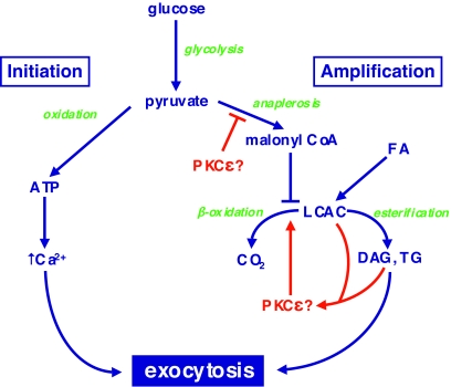
Putative site of action of PKCɛ in the amplification pathway of GSIS. Pyruvate, derived from glycolysis, undergoes two metabolic fates in mitochondria that together regulate GSIS. In the initiation pathway, it is subjected to oxidative phosphorylation to generate ATP, which leads to closure of ATP-dependent K+ channels, depolarization, and the gating of Ca2+ influx. In the amplification pathway, pyruvate augments Krebs cycle intermediates (anaplerosis), some of which can be exported to the cytosol to generate malonyl-CoA. This results in an inhibition of the β-oxidation of LCACs derived from exogenous fatty acids or mobilization of endogenous lipid stores. This favors the formation of esterification products di- or triacylglycerol (DAG or TG, respectively). PKCɛ, potentially activated by LCACs or DAG, appears to promote oxidation of lipid fuels at the expense of esterification pathways that are implicated in the amplification pathway. Whether PKCɛ acts directly at this site, or upstream at a step in anaplerosis, remains to be determined. For further explanation, see the text and Ref. .
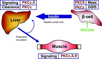
Potential sites at which individual PKC isoforms might be beneficially targeted for the treatment of type 2 diabetes. Inhibition of PKCɛ is predicted to improve insulin availability by restoring defective GSIS and by diminishing hepatic insulin clearance. This may also improve insulin sensitivity in liver and muscle. Targeting PKCδ may be of benefit in maintaining β-cell mass and in treating insulin resistance in muscle and liver. See text for details.
References
-
- Farese RV: Phospholipid signaling systems in insulin action. Am J Med 85 :36 –43,1988 - PubMed
-
- Akimoto K, Mizuno K, Osada S, Hirai S, Tanuma S, Suzuki K, Ohno S: A new member of the third class in the protein kinase C family, PKCλ, expressed dominantly in an undifferentiated mouse embryonal carcinoma cell line and also in many tissues and cells. J Biol Chem 269 :12677 –12683,1994 - PubMed
-
- Selbie LA, Schmitz-Peiffer C, Sheng YH, Biden TJ: Molecular cloning and characterization of PKCι, an atypical isoform of protein kinase-C derived from insulin-secreting cells. J Biol Chem 268 :24296 –24302,1993 - PubMed
Publication types
MeSH terms
Substances
LinkOut - more resources
Full Text Sources
Other Literature Sources
Medical
