

doi: 10.1128/JB.00155-08.
Epub 2008 Jun 27.
Loss of flagellum-based motility by Listeria monocytogenes results in formation of hyperbiofilms
Affiliations
- PMID: 18586947
- PMCID: PMC2519545
- DOI: 10.1128/JB.00155-08
Item in Clipboard
Loss of flagellum-based motility by Listeria monocytogenes results in formation of hyperbiofilms
J Bacteriol.
2008 Sep.
Abstract
Biofilm formation by the gram-positive, motile, food-borne pathogen Listeria monocytogenes was demonstrated to occur by an ordered series of stages. Biofilm development involves flagellum-based motility, which when blocked decreases initial bacterial surface attachment but subsequently leads to the formation of hyperbiofilms, surface-attached communities reaching high density.
Figures
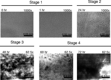
Bright-field microscopy of flow cells inoculated with L. monocytogenes 10403s. The flow cell at ambient temperature was inoculated and allowed to remain static for 1 h. Medium flow was then restored, and images of surface-attached cells were captured at a magnification of either ×1,000 to image individual surface-attached cells or ×62.5 to visualize masses of surface-attached cells (dark regions against light background). The time points and level of magnification for each image are indicted in the upper left and right, respectively. Stage 1, surface attachment of individual cells; stage 2, microcolony formation; stage 3, biofilm maturation; stage 4, community dissociation (60 h) followed by regeneration (72 h).
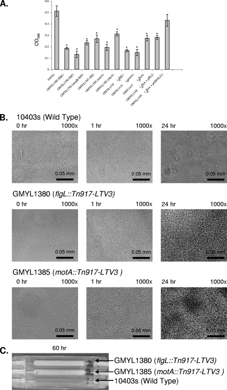
Loss of flagellar motility alters the dynamics of L. monocytogenes biofilm development. (A) Quantification of crystal violet staining (optical density at 595 nm [OD595]) to indirectly measure the amount of surface-attached biomass for selected strains following 24 h of cultivation in static 96-well polyvinyl chloride microtiter plates at 30°C. Each strain examined is indicated along with the relevant genotype. Each assay was completed in triplicate. The data were statistically analyzed by a two-tailed t test to compare each mutant to the wild-type strain. Significant differences are indicated by an asterisk when P is <0.05. (B) Examination of motility mutants cultured in a flow cell at ambient temperature. Mutants display an initial lag in surface attachment (compare at 0 h). Subsequently, the mutants progressively form surface-attached communities to a greater extent than the wild-type strain. The flow cells were treated as described in the legend of Fig. 1. The time points and level of magnification for each image are indicted in the upper left and right, respectively. (C) Motility mutants form HBs, an accumulation of surface-attached cells that impede the flow of medium to the flow cell. Bacterial strains were cultivated as described in the legend of Fig. 1 but were allowed to grow for 60 h. The image was captured with a standard digital camera to show a macroscopic view. Each chamber of the flow cell shown was inoculated with a different L. monocytogenes strain as indicated.
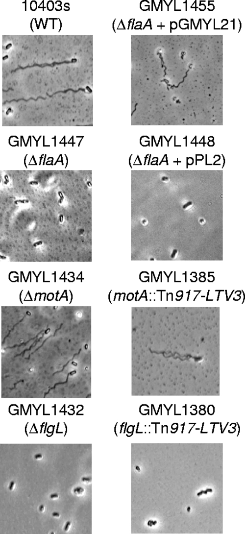
Microscopic examination of selected motility mutants and the wild-type strain. Each strain was cultivated in liquid medium as static planktonic cultures. Bacteria were stained as previously described (8). As expected, GMY1447, GMYL1448, GMY1432, and GMY1380 did not produce flagella. The wild-type (WT) strain 10403s and GMYL1455 produced flagella and were motile. Strain characteristics are indicated on the figure; plasmid pGMY21 is a flaA+ derivative of the integration vector pPL2 (10). Strains GMYL1434 and GMYL1385 produced flagella but were not motile, which is consistent with loss of motor function resulting in paralyzed flagella.
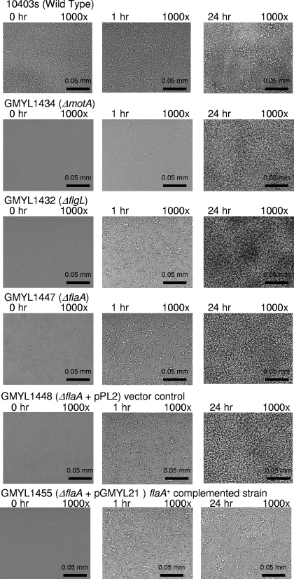
Deletion of flagellar genes affects biofilm development. The experimental conditions are as described in the legend of Fig. 1. Each chamber of the flow cell was inoculated with a different L. monocytogenes strain, as indicated above the images.
Similar articles
-
Flagellar motility is critical for Listeria monocytogenes biofilm formation.J Bacteriol. 2007 Jun;189(12):4418-24. doi: 10.1128/JB.01967-06. Epub 2007 Apr 6. J Bacteriol. 2007. PMID: 17416647 Free PMC article.
-
Listeria monocytogenes flagella are used for motility, not as adhesins, to increase host cell invasion.Infect Immun. 2006 Dec;74(12):6675-81. doi: 10.1128/IAI.00886-06. Epub 2006 Sep 18. Infect Immun. 2006. PMID: 16982842 Free PMC article.
-
Molecular biology of surface colonization by Listeria monocytogenes: an additional facet of an opportunistic Gram-positive foodborne pathogen.Environ Microbiol. 2011 Apr;13(4):835-50. doi: 10.1111/j.1462-2920.2010.02378.x. Epub 2010 Nov 18. Environ Microbiol. 2011. PMID: 21087384 Review.
-
Effect of flagella on initial attachment of Listeria monocytogenes to stainless steel.Appl Environ Microbiol. 2000 Feb;66(2):860-3. doi: 10.1128/AEM.66.2.860-863.2000. Appl Environ Microbiol. 2000. PMID: 10653766 Free PMC article.
-
Current knowledge and perspectives on biofilm formation: the case of Listeria monocytogenes.Appl Microbiol Biotechnol. 2013 Feb;97(3):957-68. doi: 10.1007/s00253-012-4611-1. Epub 2012 Dec 12. Appl Microbiol Biotechnol. 2013. PMID: 23233205 Review.
Cited by
-
Role of extracellular DNA during biofilm formation by Listeria monocytogenes.Appl Environ Microbiol. 2010 Apr;76(7):2271-9. doi: 10.1128/AEM.02361-09. Epub 2010 Feb 5. Appl Environ Microbiol. 2010. PMID: 20139319 Free PMC article.
-
Listeria monocytogenes Biofilm Adaptation to Different Temperatures Seen Through Shotgun Proteomics.Front Nutr. 2019 Jun 14;6:89. doi: 10.3389/fnut.2019.00089. eCollection 2019. Front Nutr. 2019. PMID: 31259174 Free PMC article.
-
Arcobacter butzleri Biofilms: Insights into the Genes Beneath Their Formation.Microorganisms. 2022 Jun 23;10(7):1280. doi: 10.3390/microorganisms10071280. Microorganisms. 2022. PMID: 35888999 Free PMC article.
-
Counterclockwise rotation of the flagellum promotes biofilm initiation in Helicobacter pylori.mBio. 2024 Jun 12;15(6):e0044024. doi: 10.1128/mbio.00440-24. Epub 2024 May 3. mBio. 2024. PMID: 38700325 Free PMC article.
-
Listeria monocytogenes Biofilms in Food-Associated Environments: A Persistent Enigma.Foods. 2023 Sep 6;12(18):3339. doi: 10.3390/foods12183339. Foods. 2023. PMID: 37761048 Free PMC article. Review.
References
-
- Chae, M. S. 2001. Cell viability of Listeria monocytogenes biofilms. Food Microbiol. 18103-112.
-
- Chae, M. S., and H. Schraft. 2000. Comparative evaluation of adhesion and biofilm formation of different Listeria monocytogenes strains. Int. J. Food Microbiol. 62103-111. - PubMed
Publication types
MeSH terms
Substances
LinkOut - more resources
Full Text Sources
