. 2008 Sep;295(3):L497-504.
doi: 10.1152/ajplung.90210.2008. Epub 2008 Jun 27.

Activation of AMPK attenuates neutrophil proinflammatory activity and decreases the severity of acute lung injury

Affiliations

Activation of AMPK attenuates neutrophil proinflammatory activity and decreases the severity of acute lung injury

Xia Zhao et al. Am J Physiol Lung Cell Mol Physiol. 2008 Sep.

Abstract

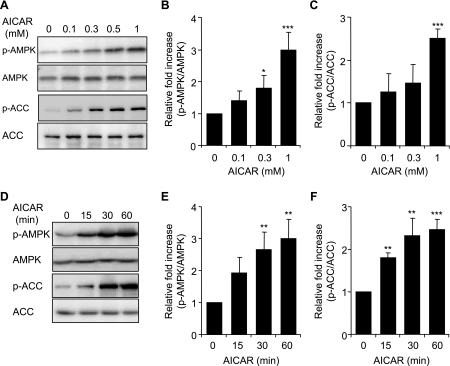
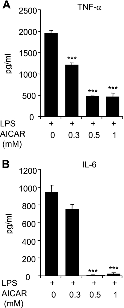
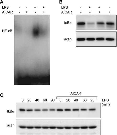
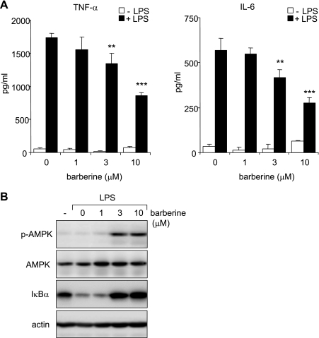
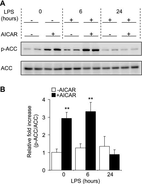
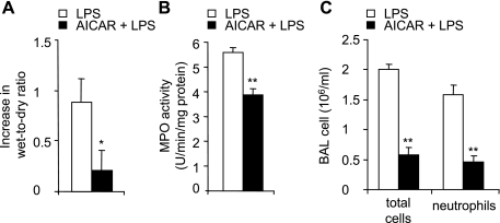
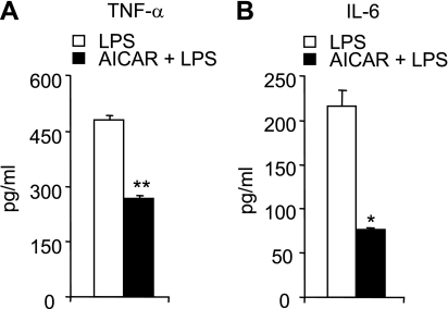
AMP-activated protein kinase (AMPK) is activated by increases in the intracellular AMP-to-ATP ratio and plays a central role in cellular responses to metabolic stress. Although activation of AMPK has been shown to have anti-inflammatory effects, there is little information concerning the role that AMPK may play in modulating neutrophil function and neutrophil-dependent inflammatory events, such as acute lung injury. To examine these issues, we determined the effects of pharmacological activators of AMPK, 5-aminoimidazole-4-carboxamide-1-beta-d-ribofuranoside (AICAR) and barberine, on Toll-like receptor 4 (TLR4)-induced neutrophil activation. AICAR and barberine dose-dependently activated AMPK in murine bone marrow neutrophils. Exposure of LPS-stimulated neutrophils to AICAR or barberine inhibited release of TNF-alpha and IL-6, as well as degradation of IkappaBalpha and nuclear translocation of NF-kappaB, compared with findings in neutrophil cultures that contained LPS without AICAR or barberine. Administration of AICAR to mice resulted in activation of AMPK in the lungs and was associated with decreased severity of LPS-induced lung injury, as determined by diminished neutrophil accumulation in the lungs, reduced interstitial pulmonary edema, and diminished levels of TNF-alpha and IL-6 in bronchoalveolar lavage fluid. These results suggest that AMPK activation reduces TLR4-induced neutrophil activation and diminishes the severity of neutrophil-driven proinflammatory processes, including acute lung injury.


Figures

Fig. 1.
Fig. 1.
Effects of 5-aminoimidazole-4-carboxamide-1-β-d-ribofuranoside (AICAR) on AMP-activated protein kinase (AMPK) and acetyl-CoA carboxylase (ACC) phosphorylation (p-) in bone marrow neutrophils. Neutrophils were cultured with AICAR (0, 0.1, 0.3, or 1 mM) for 60 min (AC) or with 1 mM AICAR for the indicated time periods (DF). Cell lysates were subjected to SDS-PAGE and Western blot analysis using antibodies specific for phospho-AMPK (Thr172), total AMPK, phospho-ACC (Ser79), and total ACC. A and D show representative Western blots, whereas B, C, E, and F demonstrate mean band optical density obtained from 3 individual experiments. Data are expressed as p-AMPK/AMPK or p-ACC/ACC ratios. Means ± SE, n = 3; *P < 0.05; **P < 0.01; ***P < 0.001 compared with control (untreated cells).
Fig. 2.
Fig. 2.
AICAR inhibits LPS-induced TNF-α and IL-6 production by neutrophils. Cells were treated with the indicated doses of AICAR for 1 h followed by LPS (0 or 100 ng/ml) for 4 h. Levels of TNF-α (A) and IL-6 (B) in the culture supernatants were determined using ELISA assays. Values are expressed as means ± SE using results from 3 independent experiments. ***P < 0.001 compared with LPS-treated cells.
Fig. 3.
Fig. 3.
AMPK inhibits LPS-induced IκBα degradation and NF-κB nuclear accumulation in LPS-treated neutrophils. Neutrophils were treated with AICAR (0 or 1 mM) for 1 h followed by LPS (0 or 100 ng/ml) for 1 h. A: representative EMSA showing inhibitory effects of AICAR on LPS-induced nuclear accumulation of NF-κB. B: IκBα levels in LPS-stimulated neutrophils treated or untreated with AICAR. C: neutrophils were pretreated with AICAR (0 or 1 mM) for 1 h, and degradation of IκBα was determined after at baseline and 20, 40, 60, and 90 min after the addition of LPS (100 ng/ml) to the cultures. A representative Western blot of IκBα and actin are shown. A 2nd experiment with neutrophils derived from a separate group of mice provided similar results.
Fig. 4.
Fig. 4.
Barberine-dependent activation of AMPK inhibits LPS-induced IκBα degradation and cytokine production. A: neutrophils were cultured with barberine (0, 1, 3, or 10 μM) for 60 min followed by stimulation with LPS (0 or 100 ng/ml) for 4 h. Levels of TNF-α and IL-6 in the culture media were measured using ELISA assays. Means ± SE, n = 3; ***P < 0.001 or **P < 0.01 compared with LPS-treated cells. B: representative Western blots show levels of phospho-AMPK (Thr172), total AMPK, IκBα, or actin in cell extracts obtained from neutrophils treated with barberine (0, 1, 3, or 10 μM) for 60 min and then LPS (100 ng/ml) for an additional 1 h. A 2nd experiment with neutrophils obtained from a separate group of mice provided similar results.
Fig. 5.
Fig. 5.
Effects of AICAR on ACC phosphorylation in vivo. Mice were injected intraperitoneally with saline or AICAR (500 mg/kg) in saline 4 h before intratracheal PBS or PBS with LPS (1 mg/kg) administration. Lung homogenates were collected in mice treated with AICAR or saline 4 h previously or 6 or 24 h after LPS treatment, and samples were subjected to Western blot analysis using anti-phosphorylated-ACC (Ser79) and anti-total ACC specific antibodies. A: representative Western blots. B: quantitative densitometry of p-ACC/ACC ratios (means ± SE) with n = 3 mice/group. **P < 0.01 comparing control (LPS/PBS) to LPS/AICAR-treated mice.
Fig. 6.
Fig. 6.
AICAR attenuates LPS-induced acute lung injury in vivo. Mice were treated intratracheally with either PBS or PBS with LPS (1 mg/kg) or were given intraperitoneal saline or saline with AICAR (500 mg/kg) followed 4 h later with either PBS or PBS with LPS (1 mg/kg) intratracheally. Lungs and bronchoalveolar lavage (BAL) were obtained 24 h after LPS administration. A: effects of AICAR treatment on LPS-induced increase in lung wet-to-dry ratios. B: lung MPO levels. C: number of total white cells and neutrophils in BAL fluid. Means ± SE are shown using results from n = 6 mice per group; *P < 0.05 or **P < 0.01 when comparing AICAR/LPS to mice treated with LPS alone.
Fig. 7.
Fig. 7.
AICAR decreases LPS-induced pulmonary cytokine levels in vivo. BAL levels of TNF-α (A) and IL-6 (B). BAL fluid was obtained 24 h after LPS administration in mice treated intraperitoneally with either saline or saline with AICAR (500 mg/kg) 4 h before intratracheal LPS. Means ± SE are shown. n = 6 mice in each group; *P < 0.05 and **P < 0.01 comparing the AICAR/LPS group to mice treated with PBS/LPS.

References

    1. Abraham E Alterations in cell signaling in sepsis. Clin Infect Dis 41, Suppl 7: S459–S464, 2005. - PubMed
    1. Abraham E Neutrophils and acute lung injury. Crit Care Med 31: S195–S199, 2003. - PubMed
    1. Abraham E NF-kappaB activation. Crit Care Med 28: N100–N104, 2000. - PubMed
    1. Abraham E, Arcaroli J, Carmody A, Wang H, Tracey KJ. HMG-1 as a mediator of acute lung inflammation. J Immunol 165: 2950–2954, 2000. - PubMed
    1. Abraham E, Carmody A, Shenkar R, Arcaroli J. Neutrophils as early immunologic effectors in hemorrhage- or endotoxemia-induced acute lung injury. Am J Physiol Lung Cell Mol Physiol 279: L1137–L1145, 2000. - PubMed

Publication types

MeSH terms

LinkOut - more resources