. 2008 Oct;22(10):3628-37.
doi: 10.1096/fj.08-108274. Epub 2008 Jun 27.

Engagement of Toll-like receptor-2 on cytotoxic T-lymphocytes occurs in vivo and augments antitumor activity

Affiliations

Engagement of Toll-like receptor-2 on cytotoxic T-lymphocytes occurs in vivo and augments antitumor activity

Nicole Asprodites et al. FASEB J. 2008 Oct.

Abstract

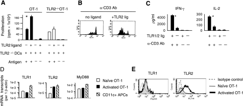
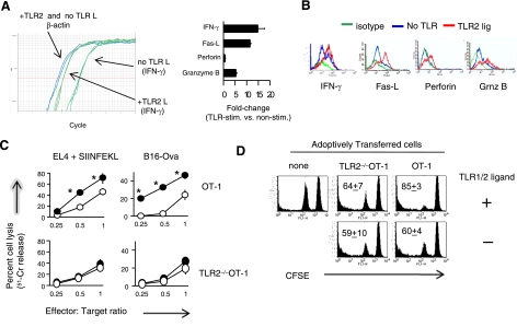
Toll-like receptors (TLRs) are among the fundamental molecules that alert the immune system to the presence of an infection by recognizing pathogen-associated molecules. Much of our understanding regarding TLR function stems from the study of innate immune cells. Recent studies by several groups, including ours, have shown that TLRs can function as costimulatory receptors for antigen-specific T cells, resulting in enhanced T-cell survival and increased expression of effector molecules. We report that the ligation of the TLR1/2 heterodimer on OT-1 cytotoxic T-lymphocytes (CTL) but not TLR2(-/-)OT-1 T cells increased cytolytic activity in vitro and in vivo. On the basis of these data, we tested the hypothesis that TLR1/2 stimulation on CTLs would enhance antitumor activity in a therapeutic model of B16-Ova melanoma. Adoptive OT-1 T-cell transfer into wild-type and MyD88(-/-) mice, followed by injection with TLR1/2 ligand, resulted in a synergistic antitumor effect, which correlated with the induction of CD8 T cells specific to various tumor antigens. In contrast, mice receiving TLR2(-/-)OT-1 T cells and TLR1/2 ligand showed minimal therapeutic efficacy. These findings emphasize the physiological significance of TLR2 engagement on CTLs and could make possible new approaches for the development of effective immunotherapies by manipulating TLR signaling within CTLs.


Figures

Figure 1.
Figure 1.
TLR1/2 engagement costimulates CTL responses. A) OT-1 T cells were purified by negative selection followed by positive selection using magnetic beads. OT-1 cells (3×10⁴) were stimulated in vitro with 3 × 10³ antigen-pulsed (SIINFEKL) H2-Kb TLR2–/– DCs in the presence or absence of 10 μg/ml TLR1/2 ligand. T-cell proliferation was measured at the end of 5 days. Values represent average ± sd of replicate samples. B) OT-1 T cells were labeled with CFSE and stimulated with plate-bound anti-CD3 antibody (1.0 μg/ml) for 5 days in the presence or absence of TLR1/2 ligand (10 μg/ml). C) OT-1 T cells were activated with-plate bound anti-CD3 (2.5 μg/ml) antibody for 72 h in the absence or presence of TLR1/2 ligand. The amount of cytokines secreted by cells was determined by ELISA. D) After 24 h of activation (black bar) or not (white bar), T cells were purified by positive selection using magnetic beads, and the expression of the indicated mRNA transcripts was determined using RT-PCR. For control, CD11+ APCs were sorted by magnetic beads (slashed bar) from spleens of WT mice and the level of transcripts was determined via RT-PCR. E) Alternatively, the surface expression of TLR1 and TLR2 proteins on nonactivated (gray fill) or activated OT-1 T cells (bold line) was evaluated using flow cytometry. All data are representative of at least 3 independent experiments, each yielding similar results. *P < 0.05; Student’s t test.
Figure 2.
Figure 2.
TLR1/2 engagement on OT-1 CTLs enhances the expression of effector molecules and augments cytolytic function. A) OT-1 T cells were purified and stimulated with peptide-pulsed TLR2–/– DCs, in the absence or presence of TLR1/2 ligand. After 24 h of activation, T cells were sorted, and the fold changes in gene expression in TLR1/2-stimulated and non-TLR-stimulated cells were determined using RT-PCR. B) Alternatively, the intracellular levels of granzyme-B, perforin, IFN-γ, and the surface expression of Fas ligand were determined by flow cytometry 72 h after activation. C) Purified OT-1 T cells were stimulated with peptide-pulsed TLR2–/– DCs in the absence (open circles) or presence (filled circles) of TLR1/2 ligand. After 5 days, OT-1 and TLR2–/–OT-1 T-cell cytoxicity was tested against peptide-pulsed EL4 or B16-Ova target cells at the indicated effector to target ratios using a standard 4-h 51Cr-release assay. D) OT-1 T cells were stimulated in vitro for 48 h and injected (i.v.) into naive MyD88–/– mice (n=3) 2 days later. Five days after AT, MyD88–/– splenocytes (target cells) were labeled with CFSEhigh and pulsed with an irrelevant H-2Kb-restricted CTL epitope (VVYDFLKC), and the other group was labeled with CFSElow and pulsed with the Ova257–264 (SIINFEKL) peptide. CFSEhigh and CFSElow target cells were mixed at a 1:1 ratio and injected (i.v.) with or without TLR1/2 agonist. The percentage of target cells was determined by flow cytometry. Graphs show average ± sd percentage lysis; 3 mice/group. Data in A, B are representative of 3 independent experiments; data in C, D are representative of 2 experiments, each showing identical trends. *P ≤ 0.02; ANOVA.
Figure 3.
Figure 3.
TLR1/2 engagement on OT-1 T cells in vivo enhances antitumor activity against established B16-Ova melanoma tumors. A) Model depicting experimental design. B–G) WT (B–D) or MyD88–/– mice (E–G) were injected (s.c.) with 25 × 103 B16-Ova tumor cells. When tumors reached a size of 5 mm2, mice were injected (i.v.) with 2.5 × 10⁶ OT-1 or TLR2–/–OT-1 T cells. Prior to AT, CTLs were activated in vitro for 48 h, as described in Materials and Methods. Mice were injected peritumorally with TLR1/2 ligand (bold line) or PBS (light line) every 5 days, starting on the same day that mice received T cells. Tumor sizes (mm2) were calculated by measuring perpendicular by longitudinal diameter. Data are compiled from 3 independent experiments. Each experiment yielded similar trends. Statistical analyses were performed using the MIXED procedure of SAS v9.13, and tumor volume kinetics were assessed by fitting quadratic random coefficient models; *P < 0.001.
Figure 4.
Figure 4.
TLR1/2-engaged CTLs promote antigen-cross presentation. Graphs are representative dot plots of tetramer-stained CD8 T cells obtained from mice that received OT-1, TLR2–/–OT-1, or no T cells in the absence or presence of TLR1/2 ligand, as in Fig. 3A. Mice were sacrificed or died 30–35 days after tumor challenge; percentage of Ova257–264-, Trp-2-, or mouse gp100-specific CD8+ T cells in the draining lymph nodes was determined by staining cells with anti-CD8 antibody and the different tetramers or pentamers. Data represent average ± sd percentage of tetramer-positive CD8 T cells; 3–4 mice/group.
Figure 5.
Figure 5.
Endogenous CD8 T cells show cytolytic activity against B16 melanoma tumor antigens. WT mice were injected with B16-Ova tumor cells followed by AT of OT-1 or TLR2–/–OT-1 T cells, as described in Fig. 3. All mice were injected with TLR1/2 ligand every 5 days, starting on the same day that tumor-bearing mice received CTLs. Between 30 and 35 days after tumor challenge, the spleens and lymph nodes were pooled from the indicated groups. CD8 T cells were purified by negative selection. To expand tumor-specific CTLs, purified CD8 T cells were restimulated with IFN-γ-treated, irradiated B16 tumor cells for 5 days in the presence of IL-2 (100 U/ml). T-cell cytotoxicity was determined in a 4 h 51Cr-release cytotoxicity assay against the indicated target cells. All experimental determinations were performed in triplicate; averages ± sd were consistently within 15% of the mean. Error bars represent mean ± sd of triplicate samples. *P ≤ 0.03; ANOVA.

Similar articles

Cited by

References

    1. Krieg A M. Development of TLR9 agonists for cancer therapy. J Clin Invest. 2007;117:1184–1194. - PMC - PubMed
    1. Akira S, Hemmi H. Recognition of pathogen-associated molecular patterns by TLR family. Immunol Lett. 2003;85:85–95. - PubMed
    1. Krieg A M. Therapeutic potential of Toll-like receptor 9 activation. Nat Rev Drug Discov. 2006;5:471–484. - PubMed
    1. Iwasaki A, Medzhitov R. Toll-like receptor control of the adaptive immune responses. Nat Immunol. 2004;5:987–995. - PubMed
    1. Takeda K, Kaisho T, Akira S. Toll-like receptors. Annu Rev Immunol. 2003;21:335–376. - PubMed

Publication types

MeSH terms