. 2008 Jul;211(Pt 14):2263-74.
doi: 10.1242/jeb.015818.

Light and peptidergic eclosion hormone neurons stimulate a rapid eclosion response that masks circadian emergence in Drosophila

Affiliations

Light and peptidergic eclosion hormone neurons stimulate a rapid eclosion response that masks circadian emergence in Drosophila

Susan L McNabb et al. J Exp Biol. 2008 Jul.

Abstract

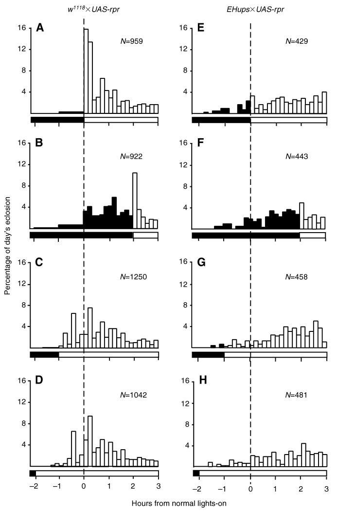
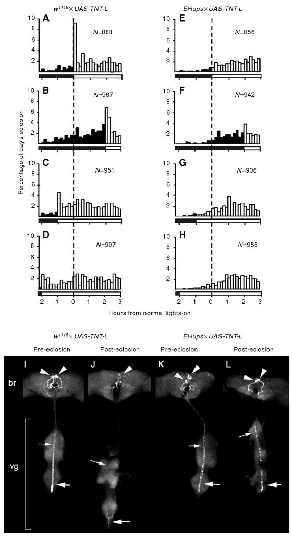
Light signals can entrain circadian clocks, but they can also mask aspects of the circadian output. We have analyzed the masking effects of a lights-on (LOn) signal on Drosophila eclosion. The LOn response results in 12-21% of the flies that emerge on a given day eclosing within 10 min of the LOn signal. Flies that lack the neuropeptide eclosion hormone (EH), or in which its release is inhibited by the tetanus toxin light chain, lack the response. Optic photoreceptors in both the ocelli and the compound eyes appear to be required for the response. The LOn signal has two effects: (1) it drastically reduces the interval between EH release and eclosion, presumably by suppressing a transient descending inhibition that immediately follows EH release, and (2) it stimulates premature EH release. The LOn signal does not influence the latency of wing spreading, an EH-regulated post-ecdysis behavior.


Figures

Fig. 1
Fig. 1
Effects of developmental age on the rapid eclosion response to light. Representative data for a population of w1118×UAS-rpr flies that resulted from a 1 day egg collection, monitored every morning for 4 days. Emerging flies were collected every 10 min between −1 h and +2 h relative to lights-on (LOn, 0 h). The amount of eclosion is normalized to the day's total eclosion. The horizontal bar below the day 4 panel represents the time relative to lights-on; black for the dark, white for the light. N, the total number of flies collected each day.
Fig. 2
Fig. 2
Effects of a light pulse on the rapid eclosion response. Flies of the w1118×UAS-rpr strain (A) received light continuously from the time of normal LOn, (B) were held in the dark or (C) received a 20 min pulse of light beginning at +1 h from normal LOn. Axes and coloring are as described for Fig. 1. Data bars give the means ± s.e.m. for four trials (N=993–1316 per test condition per trial).
Fig. 3
Fig. 3
Effects of varying the time of the LOn signal and of loss of the eclosion hormone (EH) neurons on the LOn response. Eclosion of (A–D) the w1118×UAS-rpr control flies and (E–H) EHups×UAS-rpr flies that lack EH neurons. Flies received one of four treatments: (A,E) light beginning at normal LOn, (B,F) held in the dark until +2 h, (C,G) light beginning at −1 h or (D,H) light beginning at −2 h, as represented by the horizontal bars at the base of each panel. Legends are as described for Fig. 1. Data from an individual representative experiment is shown.
Fig. 4
Fig. 4
Effects of lack of optic photoreceptors on the LOn response. The LOn shift paradigm was used to test the CS (Canton-S, control; A–D), the oc1 ocelliless (E–H) and the clieya-2 eyeless strains (I–L). Legends are as described for Fig. 3 except that, for oc1 and clieya-2, some collection bins were greater than 10 min intervals but were averaged in 10 min intervals as indicated by stippling.
Fig. 5
Fig. 5
Effects of expressing TeTxLC in the EH neurons on the LOn response and EH release. Representative results are shown for the LOn shift assay for w1118×UAS-TNT-L (A–D) and EHups×UAS-TNT-L (E-H) strains. Data are presented as described for Fig. 3. (I–L) Immunostaining for EH in CNSs from w1118×UAS-TNT-L flies (I) 8–11 h prior to eclosion and (J) within 1 min after eclosion, and from EHups×UAS-TNT-L flies (K) 8–11 h prior to eclosion and (L) within 1 min after eclosion. These are representative images of projected z-series. Arrowheads indicate the EH cell bodies located in the brain (br); large arrows, the posterior portion of the EH axon that extends down the ventral ganglion (vg); small arrows, the thoracic portion of the EH axon.
Fig. 6
Fig. 6
Effects of the LOn signal on wing spreading latency (WSL) is shown for flies that eclosed in response to LOn (upper panels, white bars) and flies that eclosed in the dark (lower panels, black bars) for (A) w1118×UAS-rpr, (B) EHups×UAS-rpr, (C) w1118×UAS-TNT-L, (D) EHups×UAS-TNT-L, (E) Canton-S, (F) oc1 and (G) and clieya-2 strains. The WSL is expressed per 10 min bin. The average WSL is indicated by an arrow.
Fig. 7
Fig. 7
Effects of light on EH release in w1118×UAS-rpr flies. Pharate adults that were not destined to eclose at the time of normal LOn were collected the night prior to eclosion and their CNSs were harvested (A,E) the night prior to eclosion, (B,F) 1 h before normal LOn, (C,G) 10–20 min after LOn or (D,H) held in the dark until 10–20 min after normal LOn. (A–D) CNSs immunostained for EH were scored for level of staining. (E–H) Representative Z projections.
Fig. 8
Fig. 8
Light stimulates rapid eclosion in competent flies, but also appears to have a second, delayed, effect. (A) Data from the w1118×UAS-rpr pulse experiment (Fig. 2B,C) show that the proportion of flies that eclosed during the first 10 min of the 20 min light pulse (white bars) was roughly equivalent to the proportion that eclosed over the corresponding 60 min interval in the dark (bracketed by dashed lines). (B) Summary of the effects of light on eclosion, adapted from the w1118×UAS-rpr pulse experiment (Fig. 2C). Dashed arrows indicate the intervals over which flies that have released EH are recruited to eclose by a LOn signal. Pharate adults that had released EH and were competent to eclose within approximately 60 min were recruited to eclose in the first 10 min after light exposure (gray arrow). If the pool of competent flies all eclosed at or shortly after LOn, the amount of eclosion after the light pulse would be expected to diminish to 0 (thick black line). Instead, the proportion of flies eclosing went down to only approximately 2% (thin black line) possibly as a result of light stimulating EH release in a group that was developmentally mature and ready to eclose.
Fig. 9
Fig. 9
A model of effects of light on eclosion. EH activates both an eclosion activation pathway and a set of inhibitory neurons that repress eclosion behavior. The activation pathway may include the CCAP neurons and additional EH-downstream neurons. We postulate that the release of CCAP and other eclosion activators is inhibited by EH action. Light suppresses the inhibitory pathway to allow the release of CCAP and other factors and permit subsequent eclosion. In addition, light acts on the EH cells to stimulate EH release, probably via the retinal photoreceptors. Although eclosion can be accelerated by light, wing spreading cannot, suggesting that EH stimulates these behaviors via distinct pathways. Arrow and line thickness indicate the strengths of the different responses.

References

    1. Aschoff J. Exogenous and endogenous components in circadian rhythms. Cold Spring Harbor Symp Quant Biol. 1960;25:11–28. - PubMed
    1. Ashburner M. Drosophila: A Laboratory Manual. Cold Spring Harbor, NY: Cold Spring Harbor Laboratory Press; 1989.
    1. Baker JW, McNabb SL, Truman JW. The hormonal coordination of behavior and physiology at adult ecdysis in Drosophila melanogaster. J Exp Biol. 1999;202:3037–3048. - PubMed
    1. Benzer S. Behavioral mutants of Drosophila isolated by countercurrent distribution. Proc Natl Acad Sci USA. 1967;58:1112–1119. - PMC - PubMed
    1. Binkley S, Mosher K, Reilly K. Circadian rhythms in house sparrows: Lighting ad lib. Physiol Behav. 1983;31:829–893. - PubMed

Publication types

MeSH terms

LinkOut - more resources